
Paranasal sinus mucoceles
Mucoceles, surgery
Authors
Abstract
Mucoceles of the paranasal sinus are benign expanding lesions that primarily occur between the fourth and seventh decades of life. Sinus mucoceles may develop due to the obstruction of the normal passageway by trauma, inflammation, mass lesions, idiopathic, or iatrogenic causes. The most common site for the evolution of a mucocele is within the frontal sinuses, followed in frequency by the ethmoidal, maxillary, and sphenoid sinuses. The symptoms depend on the location and size of the mucocele. The symptoms can be classified as nasal, ophthalmologic, intracranial, and cosmetic. A mucocele can become infected, forming a mucopyocele, with a risk of infectious complications including meningitis, orbital cellulitis, and osteomyelitis. Today, the first choice of treatment is marsupialization of the mucoceles using endoscopic sinus surgery. External approaches to frontal sinus are still used, in erosion of the anterior frontal sinus wall and far lateral located lesions.
Keywords
The paranasal sinus mucocele is a dilated mucous-filled sinus that is lined by mucous membranes.1 Mucoceles of the paranasal sinus are benign, expanding lesions that primarily occur between the fourth and seventh decades of life. The incidence is similar for both genders.2,3,4 Paranasal sinus mucoceles are extremely rare in children and adolescents. Cystic fibrosis is the main etiologic factor.5 The symptoms depend on the location and size of the mucocele. The mechanism underlying mucocele formation is multifactorial. Impediments to sinus ostium ventilation are thought to be the principal cause. Sinus mucoceles may develop due to the obstruction of the normal passageway by trauma, inflammation, mass lesions, idiopathic, or iatrogenic causes 6,7,8 (Figure 1). However, the predisposing etiology of primary and secondary nasal mucoceles seems to be different. For the formation of primary mucoceles, inflammatory blockages of mucous drainage, secretory duct obstruction, cystic dilation of mucosal glands, and cystic degeneration of polyps were proposed as possible causes. On the other hand, secondary mucoceles sequestering residual mucosa in the wound and long-term retention of tissue fluid have also been advocated as factors leading to their formation.9,10 Bockmühl U et al. has evaluated 290 patients with sinus mucocele according to their etiologies. They reported incidences related to: a previous operation 66% (168 patients), trauma 14% (37 patients), chronic sinusitis 2% (5 patients), tumor 1% (2 patients), and unknown causes 17% (43 patients). When mucoceles due to a previous operation were further evaluated, it was concluded that 98% were due to external procedures such as Jansen-Ritter and Caldwell-Luc , and 2% were due to endonasal surgery. On average, the periods of time between external surgery and occurrence were as follows: maxillary sinus 15 years, fronto-ethmoid 13 years, ethmoid sinus 10 years, and frontal and sphenoid sinus 8 years.11,12 It is reported that the use of alloplastic materials in the management of frontal sinus fractures may be a cause of orbital mucopyocele.13Mucoceles grow slowly. As pressure builds due to continuous swelling, the surrounding bony structures begin to thin, contributing to further resorption and remodeling of the bone. Some authors suggest that release of osteolytic factors may contribute to the progressive development of mucoceles or mucopyoceles. Prostaglandins and collagenases aid in osteolysis and further enhance the expansile nature of the cysts.14,15,16 Lund and Milroy proposed that the obstruction to sinus outflow in combination with superimposed infection caused the release of cytokines from lymphocytes and monocytes. The cytokine release stimulates fibroblasts to secrete prostoglandins and collagenases, which in turn could stimulate bone resorption leading to expansion of the mucocele.17
Histopathologically, paranasal sinus mucoceles have features of respiratory mucosa with cyst walls demonstrating single-layered, pseudostratified, ciliated, columnar epithelium.15,17,18 Although metaplastic changes are rare, chronic cases show evidence of squamous metaplasia. Reactive bone formation is also possible in areas adjacent to cyst epithelium.15 Mucoceles show elevated expression of IL-12 which secondarily leads to increased expression of IL-2 and interferon gamma. Subsequently, an increased activation of TH2 lymphocytes hastens the presence of a chronic inflammatory infiltrate.16 Additional findings include cholesterol granuloma, hemorrhage, fibrosis, and granulation tissue.14 Central nervous tissue may also be identified in those cases of intracranial herniation.15 In some cases, ruptured salivary ducts may be identified. The adjacent minor salivary glands often contain a chronic inflammatory cell infiltrate and dilated ducts. Most mucoceles are self-limiting. They rupture and resolve on their own. However, other lesions are chronic in nature and local surgical excision is necessary. To minimize the risk of recurrence, when the area is excised, the surgeon should remove any adjacent salivary glands that may be supplying the lesion. The specimen should be submitted for pathologic examination to confirm the diagnosis and to rule out the possibility of a salivary gland malignancy. The prognosis is excellent, although occasionally mucoceles recur, necessitating reexcision, especially if the associated glands had not been removed.18 Radiographically, the sinus is initially opacified by retained secretions, which displaces all the air content. Pressure erosion of the surrounding bony walls by the mucocele decalcifies the mucoperiosteal cortical plate and destroys the normal scalloped borders of the sinus (Figure 2). With progression of the lesion, the sinus appears more radiolucent radiographically. There may be a zone of reactive osteitis and bone sclerosis around the margins of the mucocele. In approximately 5% of mucoceles, peripheral calcification is evident radiographically. This calcification may be large enough to stimulate the growth of an osteoma. With extensive intracranial extension by a frontal mucocele, a segment of the posterior wall of the frontal sinus may be displaced as a bony fragment deep into the frontal lobe. A view of the optic foramen may be valuable in the diagnosis of a mucocele of the frontal sinus as well as of the posterior ethmoidal cells. The sphenoid mucocele presents radiographically as a destructive expanding cystic or mass lesion.When expansion and bone destruction are present, differential diagnosis includes benign and malignant lesions of the paranasal sinuses. Benign lesions include neurofibroma; dermoid, epidermoid, cementifying fibroma, angiofibroma, inverting papilloma, and cylindrinoma. Malignant lesions include adenoid cystic carcinoma, plasmocytoma, embryonal rhabdomyosarcoma, lymphoma, schwannoma, and tumors of dental origin.18,19,20 In the absence of bone erosion, mucoceles must be differentiated from several conditions including retention cysts, chronic sinusitis, antrochoanal polyp, and polyposis of the paranasal cavities.21,22,23
A mucocele can become infected, forming a mucopyocele with the risk of infectious complications including meningitis, orbital cellulitis, and osteomyelitis. When the mucocele expands so that the sinus volume is insufficient to house the mucocele, pain and pressure erosion can occur.24 Pressure erosion can result in significant anatomic deformity including facial asymmetry and dynamic proptosis of the orbits.25
Fortunately, after the resolution of the mucocele, the deformity usually remodels. Symptoms vary depending on the location of the mucocele and may be classified as rhinological, neurologic, or most frequently, ophthalmologic.26,27 The most common site for the evolution of a mucocele is within the frontal sinuses.23,28 Sphenoid sinus mucocele is a rare condition which represents only 1-2% of all paranasal sinus mucoceles.9 In a study of 108 patients, Har-El G reported paranasal sinus mucocele occurrence rates as: frontal and fronto-ethmoid 66 patients (61%), ethmoid sinus 23 patients (22%), sphenoid sinus 12 patients (11%), and maxillary sinus 6 patients (6%).28 Caylakli et al. determined that maxillary sinus mucoceles make up 10% of all mucoceles.22
Fronto-ethmoid MucocelesMucoceles arising from the frontal sinus present with a variety of clinical signs such as decreased visual acuity, visual field abnormalities, proptosis, ptosis, periorbital swelling, displacement of the globe, restricted ocular movements and choroidal folds. The patient’s visual activity is usually not affected unless compression of the optic nerve
occurs from the mass effect of the mucocele. Meningitis, meningoencephalitis, pneumocephalus, brain abscess, seizures, and cerebrospinal fluid fistulas are other serious sequelae. The most common presenting symptom was proptosis in 61.5% of the patients reviewed. Forehead swelling and nasal congestion were each present in 38.5% of patients.
Other presenting symptoms include orbital swelling, forehead sinus cutaneous fistula, orbital cellulitis, and swelling of medial canthus.23
Possible initial sequelae include infection, cerebrospinal fluid fistula, meningitis,thrombosis, abscess, paresthesia, and headache. Less common sequelae include cosmetic abnormality, pyomucocele, intracranial abscess, headaches, and osteomylitis. Orbital involvement is rare, but includes vision loss, diplopia, superior ophthalmic vein thrombosis, cavernous sinus thrombosis, and proptosis. Sinus outflow obstruction from trauma, inflammation, previous surgery, tumors, and polyps are exacerbating factors.6 The definitive treatment of frontal mucoceles is surgical.29 Until the 1980’s, mucoceles were exenterated mainly using external approaches. Leading surgical techniques were Lynch– Howarth 30 incision and osteoplastic frontal sinusotomy as described by Bockhmul et al.31 and by Weber et al.32 Traditionally, non-endoscopic treatment of FOE mucoceles has involved trephination procedures (Figure 3) or osteoplastic flap with mucosal extirpation and fat obliteration. In 1989, Kennedy et al. proposed an alternative endoscopic approach that showed no recurrences after a mean followup of 18 months.33 Today, marsupialization of mucoceles using endoscopic sinus surgery is considered the first choice of treatment.18,28,29 It is a safe method with both low morbidity and recurrence rates (Figure 4). External approaches to frontal sinus are still used, alone or combined with ESS, but no clear-cut definition of the limits of a successful endoscopic procedure has been given.29,34,35
A survey focusing on the current management of frontal sinus disease in the United Kingdom showed that 54% of surgeons are using an external approach to treat frontalsinus mucoceles independent of their location; only 39% attempt an endoscopic drainage if the cyst lies sufficiently close to the midline.36 Correa 21 stated that patients with a narrow anterior-posterior diameter of the frontal recess, a highly comparmentalized frontal sinus, an extensive polypoid degeneration of the frontal sinus mucosa, or those with high thickened secretions are candidates for obliteration.37 Successful frontal sinus obliteration requires meticulous removal of all visible sinus mucosa and the inner cortex of the sinus wall. A wide variety of implant materials for obliteration has been described. Autologous fat transplantation is regarded as the gold standart.38 The most common reasons for choosing an osteoplastic flap procedure were erosion of the anterior frontal sinus wall requiring forehead reconstruction, far lateral located lesions, or unfavorable frontal recess anatomy with extensive scarring.23 We choose external approaches in far lateral located lesions, anterior frontal sinus wall’s defects, and huge lesions expanding into the intracranial region (Figures 5,6). On the other hand, M. Trimarchi treated frontal sinus mucoceles with lateral extension by ESS Surgical technique included anterior ethmoidectomy and either Type I, IIa or IIb frontal sinus drainage according to Draf.39,40 Eloy JA, reported the use of a balloon dilation method for drainage of a large frontal sinus mucocele (44).
Maxillary Sinus MucocelesMaxillary sinus mucoceles are more prevalent in Japan, where they are usually reported as following Caldwell-Luc maxillary sinusectomy.22,41,42 Mucoceles that develop following Caldwell-Luc operations are presumed to form as a result of entrapped sinus mucosa. Although one of the theories about development of mucocele is chronic infection, Busaba et al. compared the bacteriology of maxillary sinus mucoceles to chronic sinusitis and reported that the data does not support infection as the main origin of nontraumatic maxillary sinus mucocele.43 Caylakli et al. state that in 64% of a series of 14 patients, the etiological cause remains uncertain. The symptoms of mucoceles are related to their expansion and subsequent pressure and the obstruction of surrounding anatomic structures (Figure 7). Antral mucoceles are commonly reported to present as a painless bulging of the cheek. Medial expansion of the wall of the maxillary sinus into the nasal cavity displaces the inferior turbinate and causes the nasal obstruction.44 Superior expansion of the antrum into the inferior orbit can cause displacement of the orbital contents and visual changes. Downward displacement into the area of the alveolus can even cause a loosening of teeth.42
Retention cysts are common in the maxillary sinus and may be found on imaging studies in approximately 9% of the population. They are thought to form due to obstruction of the ducts of seromucous glands in the sinus lining, which results in an epithelium-lined cyst containing mucous or serous fluid. They develop under mucous membranes of the sinus which explains why they are so thin-walled. Radiographically, the cyst is a rounded, dome-shaped, soft tissue mass, most commonly situated on the floor of the maxillary sinus, often containing clear, yellowish fluid. Mucoceles are associated with obstruction of the duct or natural ostium of any of the paranasal sinuses and grow under the periosteum. Periosteum contributes to construction of the cystic wall, and, as a result, the wall of mucocele becomes thick and tough. The growing site of the mucocele is under the periosteum, whereas retention cysts grow under the mucosa of the sinus. This explains why retention cysts are non-expanding, well circumscribed, mucosa-covered masses, whereas mucoceles exhibit an osteolytic capacity with a tendency to expand along the path of least resistance.22,44,45
In the treatment of maxillary mucoceles, surgical techniques including external approaches, marsupialization, Caldwell-Luc procedures, and endoscopy are used.41,42,45 There was no relapse in a 14-patient series by Caylakli et al. in which endoscopic ethmoidectomy and middle meatal antrostomy were applied. We consider that an extensive marsupialization of the mucocele using middle meatal antrostomy is sufficient for treatment (Figure 8).
Sphenoid Sinus MucocelesSphenoid sinus mucocele is a rare condition which represents only 1–2% of all paranasal sinus mucoceles.46 Three pathogenetic mechanisms have been postulated for sphenoid sinus mucocele: submucosal oedema, secretory duct obstruction, and ostia obstruction. Submucosal oedema may result from allergic rhinitis causing ostia narrowing with obstruction; ostia obstruction may also be due to chronic sinusitis, polyposis, or both.47 Sphenoid sinus mucocele formation has been reported as a possible late complication of radiotherapy to the head and neck.9Radiation causes scarred mucosa which may occlude the sinus ostium.
Presenting symptoms are non-specific, and patients may be asymptomatic. The clinical symptoms and signs of sphenoid sinus mucocele are directly related to the sinus anatomy and its contiguous neurological and vascular structures. Important structures are described
which are contiguous with the sphenoid sinus: cranial nerves II to VI, cavernous sinus, carotid artery, sphenopalatine artery and nerve, pterygoid canal and nerve, dura, and pituitary gland. Patients with sphenoid sinus mucocele become symptomatic when any of these structures are displaced or compressed; mucocele commonly spreads along the path of least resistance, anteriorly, into the nasopharynx, or anterolaterally, toward the orbit. Sphenoid sinus mucocele is also more likely to present with visual changes than with inflammatory disease. Visual loss in sphenoid disease has been attributed to optic neuritis or compression.48 Hejazi et al. reviewed 130 cases of symptomatic sphenoid sinus mucocele reported up to 2001. The patients presented with these symptoms , in order of decreasing frequency: headache (87%), ocular disorders (85%), amaurosis (58%), oculomotor palsies (55%), nasal symptoms (38%), endocrine disorders (4 of 130) and anhypopituitarism (1 of 130).49 Intracranial extension is unusual in sphenoid sinus mucocele. If extension occurs, it is commonly into the suprasellar region or anterior cranial fossa. Computerized tomography provides valuable information regarding bony erosion, while MRI enables more precise visualisation of the involvement of the optic nerve, dura, pituitary gland, cavernous sinus, and carotid artery.48
Mucoceles may show various imaging features, depending on their contents. They often have low attenuation on CT, a low signal on T1-weighted MRI, and a high signal on T2-weighted MRI, due to their high water content. There is usually no enhancement, or at most marginal enhancement, on CT and T1-weighted MRI, whereas many of the lesions in the differential diagnosis show contrast enhancement. Thus, the diagnosis of mucocele can be suspected pre-operatively and differentiated from craniopharyngioma, pituitary tumor, dermoid, chordoma, epidermoid, aneurysm, metastatic lesions and, in rare cases, an arachnoid cyst. In mucoceles with a high protein content, T1-weighted MRI shows a lesion which is homogeneously hyperintense centrally with a surrounding rim of hypointense mucosa.49 Endoscopic drainage is safe and effective in eradication of disease.
Middle Concha Bullosa MucocelesA concha bullosa alone is usually asymptomatic. An enlarged concha bullosa may obstruct the middle meatus or infundibulum. Headaches, orbital pain, exophthalmia, nasal discharge, postnasal drip, nasal obstruction, and hyposmia are all possible symptoms of a concha bullosa mucopyocele. A concha bullosa mucopyocele may also manifest as migraine headaches.8
Declarations
Animal and Human Rights Statement
All procedures performed in this study were in accordance with the ethical standards of the institutional and/or national research committee and with the 1964 Helsinki declaration and its later amendments or comparable ethical standards. No animal or human studies were carried out by the authors for this article.
Data Availability
The datasets used and/or analyzed during the current study are not publicly available due to patient privacy reasons but are available from the corresponding author on reasonable request.
Conflict of Interest
None of the authors received any type of financial support that could be considered potential conflict of interest regarding the manuscript or its submission.
Funding
None.
Scientific Responsibility Statement
The authors declare that they are responsible for the article’s scientific content including study design, data collection, analysis and interpretation, writing, some of the main line, or all of the preparation and scientific review of the contents and approval of the final version of the article.
References
-
Oliverio PJ, Zinreich SJ. Radiology of the nasal cavity and paranasal sinuses. In: Cummings CW, Fredrickson JM, Harker LA, Krause CJ, Richardson MA, Schuller DE, eds. Otolaryngology: Head and Neck Surgery. 3rd ed. Missouri: Mosby; 1998:1065-1091.
-
Iannetti G, Cascone P, Valentini V, et al. Paranasal sinus mucocele: diagnosis and treatment. J Craniofac Surg. 1997;8:391-398. doi:10.1097/00001665-199708050-00011
-
Fu CH, Chang KP, Lee TJ. The difference in anatomical and invasive characteristics between primary and secondary paranasal sinus mucoceles. Otolaryngol Head Neck Surg. 2007;136(4):621-625.
-
Lai PC, Liao SL, Jou JR, et al. Transcaruncular approach for the management of frontoethmoid mucoceles. Br J Ophthalmol. 2004;88(5):725.
-
Chahed H, Romdhane N, Zainine R, et al. Pediatric mucoceles: clinical aspects and therapeutic approaches. Rev Stomatol Chir Maxillofac. 2012;113(6):437-441.
-
Shah A, Meyer DR, Parnes S. Management of frontoethmoidal mucoceles with orbital extension: is primary orbital reconstruction necessary? Ophthalmic Plast Reconstr Surg. 2007;23(4):267-271. doi:10.1097/iop.0b013e318073d19a
-
Weitzel EK, Hollier LH, Calzada G, Manolidis S. Single-stage management of complex fronto-orbital mucoceles. J Craniofac Surg. 2002;13:739-745. doi:10.1097/00001665-200211000-00004
-
Hejazi N, Witzmann A, Hassler W. Ocular manifestations of sphenoid mucoceles: clinical features and neurosurgical management of three cases and review of the literature. Surg Neurol. 2001;56:338-343.
-
Stankiewicz JA. Sphenoid sinus mucocele. Arch Otolaryngol Head Neck Surg. 1989;115:735-740.
-
Steven CM, Jorge DL, Robert HM. Mucoceles of the maxillary sinus. Otolaryngol Head Neck Surg. 1997;117:18-21.
-
Bockmühl U, Kratzsch B, Benda K, Draf W. Surgery for paranasal sinus mucocoeles: efficacy of endonasal micro-endoscopic management and long-term results of 185 patients. Rhinology. 2006;44(1):62-67.
-
Bockmühl U, Kratzsch B, Benda K, Draf W. Paranasal sinus mucoceles: surgical management and long-term results. Laryngorhinootologie. 2005;84(12):892-898.
-
Mineck CW, Chandra RK, Cohen N. Orbital mucopyocele after the use of alloplastic materials in the management of frontal sinus fractures. Otolaryngol Head Neck Surg. 2006;135(6):974-976. doi:10.1016/j.otohns.2005.09.011
-
Capra GG, Carbone PN, Mullin DP. Paranasal sinus mucocele. Head Neck Pathol. 2012;6:369-372.
-
Thompson LDR, Wenig BM. Mucocele of paranasal sinus. In: Diagnostic Pathology: Head and Neck. Salt Lake City, UT: Amirsys; 2011:45.
-
Obeso S, Llorente JL, Rodrigo JP, Sanchez R, Mancebo G, Suarez C. Paranasal sinuses mucoceles: our experience in 72 patients. Acta Otorrinolaringol Esp. 2009;60(5):332-339.
-
Lund VJ. Fronto-ethmoidal mucoceles: a histopathological analysis. J Laryngol Otol. 1991;105:921-923. doi:10.1017/s0022215100117827
-
Pierse JE, Stern A. Benign cysts and tumors of the paranasal sinuses. Oral Maxillofac Surg Clin North Am. 2012;24:249-264. doi:10.1016/j.coms.2012.01.007
-
Bell GW, Joshi BB, Macleod RI. Maxillary sinus disease: diagnosis and treatment. Br Dent J. 2011;210:113-118. doi:10.1038/sj.bdj.2011.47
-
Ng YH, Sethi DS. Isolated sphenoid sinus disease: differential diagnosis and management. Curr Opin Otolaryngol Head Neck Surg. 2011;19:16-20. doi:10.1097/moo.0b013e32834251d6
-
Tal H, Altini M, Lemmer J. Multiple mucous retention cysts of the oral mucosa. Oral Surg Oral Med Oral Pathol. 1984;58:692-695. doi:10.1016/0030-4220(84)90036-7
-
Caylakli F, Yavuz H, Cagici AC, Ozluoglu LN. Endoscopic sinus surgery for maxillary sinus mucoceles. Head Face Med. 2006;2:29. doi:10.1186/1746-160x-2-29
-
Herndon M, McMains KC, Kountakis SE. Presentation and management of extensive fronto-orbital-ethmoid mucoceles. Am J Otolaryngol. 2007;28:145-147. doi:10.1016/j.amjoto.2006.07.010
-
Chao-Jung L, Chuan-Hsiang K, Bor-Hwang K, et al. Frontal sinus mucocele presenting as oculomotor nerve palsy. Otolaryngol Head Neck Surg. 2002;126(5):588-590.
-
Malhotra R, Wormald PJ, Selva D. Bilateral dynamic proptosis due to frontoethmoidal sinus mucocele. Ophthalmic Plast Reconstr Surg. 2003;19(2):156-157. doi:10.1097/01.iop.0000055829.79494.37
-
Lee TJ, Li SP, Fu CH, et al. Extensive paranasal sinus mucoceles: a 15-year review of 82 cases. Am J Otolaryngol. 2009;30:234-238. doi:10.1016/j.amjoto.2008.06.006
-
Kim YS, Kim K, Lee JG, Yoon JH, Kim CH. Paranasal sinus mucoceles with ophthalmologic manifestations: a 17-year review of 96 cases. Am J Rhinol Allergy. 2011;25:272-275. doi:10.2500/ajra.2011.25.3624
-
Har-El G. Endoscopic management of 108 sinus mucoceles. Laryngoscope. 2001;111:2131-2134.
-
Soon SR, Lim CM, Singh H, Sethi DS. Sphenoid sinus mucocele: 10 cases and literature review. J Laryngol Otol. 2010;124:44-47. doi:10.1017/s0022215109991551
-
Trimarchi M, Bertazzoni G, Bussi M. Endoscopic treatment of frontal sinus mucoceles with lateral extension. Indian J Otolaryngol Head Neck Surg. 2013;65(2):151-156. doi:10.1007/s12070-012-0611-9
-
Bockmühl U. Osteoplastic frontal sinusotomy and reconstruction of frontal defects. In: Kountakis S, Senior B, Draf W, eds. The Frontal Sinus. Berlin: Springer; 2005:281-289. doi:10.1007/3-540-27607-6_31
-
Weber R, Draf W, Keerl R, et al. Osteoplastic frontal sinus surgery with fat obliteration: technique and long-term results using magnetic resonance imaging in 82 operations. Laryngoscope. 2000;110:1037-1044.
-
Kennedy DW, Josephson JS, Zinreich SJ, Mattox DE, Goldsmith MM. Endoscopic sinus surgery for mucoceles: a viable alternative. Laryngoscope. 1989;99:885-895. doi:10.1288/00005537-198909000-00002
-
Chiu AG, Vaughan WC. Management of the lateral frontal sinus lesion and the supraorbital cell mucocele. Am J Rhinol. 2004;18:83-86. doi:10.1177/194589240401800203
-
Yanagisawa E, Scher DA. Laterally positioned mucocele of the frontal sinus. Ear Nose Throat J. 2002;81:202-204. doi:10.1177/014556130208100404
-
Molony NC, Ah-See K, Rachmanidou A, Draf W. A survey of contemporary management of frontal sinus disease in the United Kingdom. Eur Arch Otorhinolaryngol. 2000;257(5):247-250. doi:10.1007/s004050050232
-
Correa AJ, Duncavage JA, Fortune DS, Reinisch L. Osteoplastic flap for obliteration of the frontal sinus: five years’ experience. Otolaryngol Head Neck Surg. 1999;121(6):731-735. doi:10.1053/hn.1999.v121.a98218
-
Weber R, Draf W, Keerl R, Kahle G, Schinzel S, Thomann S, et al. Osteoplastic frontal sinus surgery with fat obliteration: technique and long-term results using magnetic resonance imaging in 82 operations. Laryngoscope. 2000;110(6):1037-1044.
-
Draf W. Endonasal micro-endoscopic frontal sinus surgery: the Fulda concept. Oper Tech Otolaryngol Head Neck Surg. 1991;2:234-240. doi:10.1016/s1043-1810(10)80087-9
-
Draf W. Endonasal frontal sinus drainage type I-III according to Draf. In: Kountakis S, Senior B, Draf W, eds. The frontal sinus. Berlin: Springer; 2005:219-232. doi:10.1007/3-540-27607-6_24
-
Busaba NY, Salman SD. Maxillary sinus mucoceles: clinical presentation and long-term results of endoscopic surgical treatment. Laryngoscope. 1999;109:1446-1449. doi:10.1097/00005537-199909000-00017
-
Martin RJ, Jackman DS, Philbert RF, McCoy JM. Massive proptosis of the globe. J Oral Maxillofac Surg. 2000;58:794-799. doi:10.1053/joms.2000.7268
-
Busaba NY, Siegel N, Salman SD. Bacteriology of nontraumatic maxillary sinus mucoceles versus chronic sinusitis. Laryngoscope. 2000;110:969-971. doi:10.1097/00005537-200006000-00016
-
Khong JJ, Malhotra R, Selva D, Wormald PJ. Efficacy of endoscopic sinus surgery for paranasal sinus mucocele including modified endoscopic Lothrop procedure for frontal sinus mucocele. J Laryngol Otol. 2004;118:352-356. doi:10.1258/002221504323086534
-
Busaba NY, Kieff D. Endoscopic sinus surgery for inflammatory maxillary sinus disease. Laryngoscope. 2002;112:1378-1383. doi:10.1097/00005537-200208000-00010
-
Kosling S, Hintner M, Brandt S, Schulz T, Bloching M. Mucoceles of the sphenoid sinus. Eur J Radiol. 2004;51:1-5.
-
Hadar T, Shvero J, Nageris BI, Yaniv E. Mucus retention cyst of the maxillary sinus: the endoscopic approach. Br J Oral Maxillofac Surg. 2000;38:227-229. doi:10.1054/bjom.1999.0160
-
Rejab E, Said H, Saim L, Thim L. Sphenoid sinus mucocele: a possible late complication of radiotherapy to the head and neck. J Laryngol Otol. 1991;105:959-960. doi:10.1017/s002221510011792x
-
Sethi DS. Isolated sphenoid lesions: diagnosis and management. Otolaryngol Head Neck Surg. 1999;120:730-736. doi:10.1053/hn.1999.v120.a89436
Figures
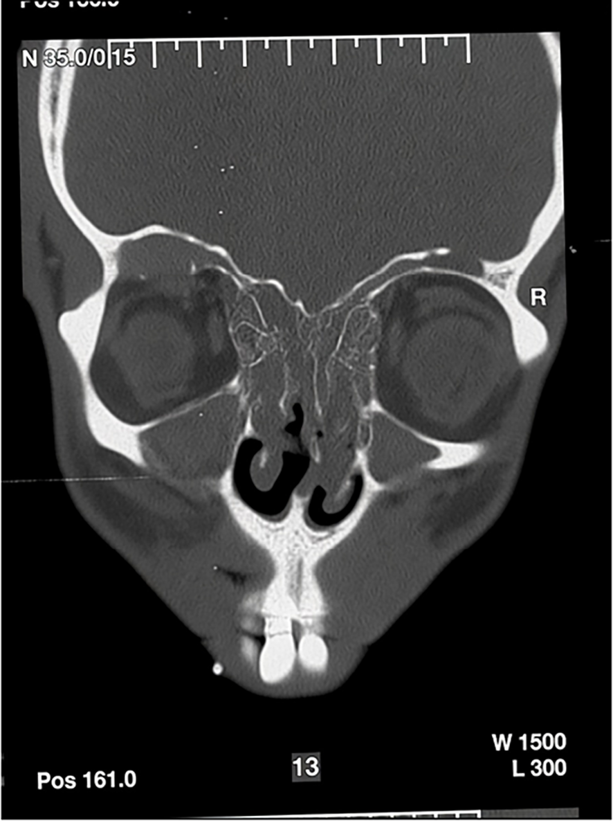
Figure 1. Frontal sinus mucocele due to nasal polyposis
Figure 2. The coronal plane CT image showing a mucocele (asteriks) in the right frontal sinus base that thinned the bone structure (A). In addition, thinning (arrows) and deformation can be observed in the right sinus roof (B) and anterior wall of the left frontal sinus (C).
Figure 3. Frontal sinus trephination in a patient that cannot be entubed due to giant hemangioma of the tongue
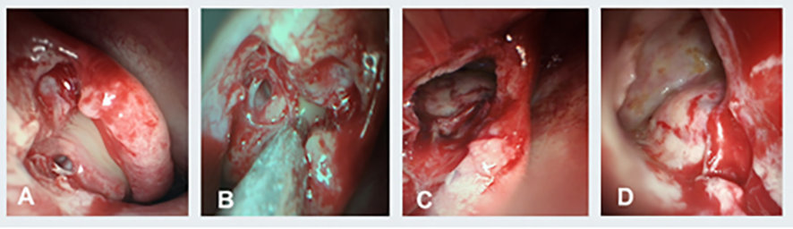
Figure 4. Endoscopic marsupialization of fronto-ethmoid mucocele
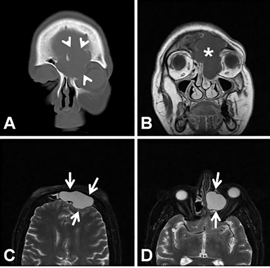
Figure 5. A huge fronto-ethmoidal located mucocele (asterisk) is shown. In the coronal CT (A) and T1 (B) and T2-weighted MR images (C, D) the expansile mucocele (arrows) causes thinning (arrowheads) of the frontal sinus walls.
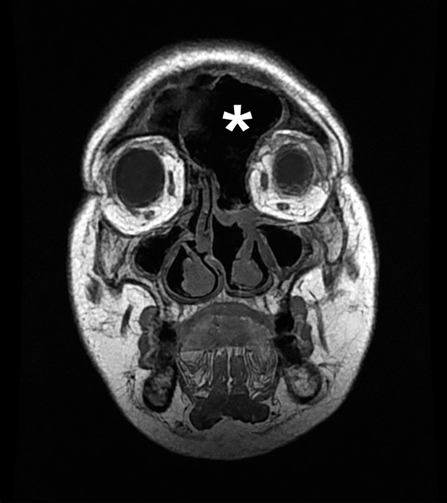
Figure 6. The coronal T1-weighted MR image after surgery (E). Air (asterisk) is observed in the left fronto-ethmoidal cavity
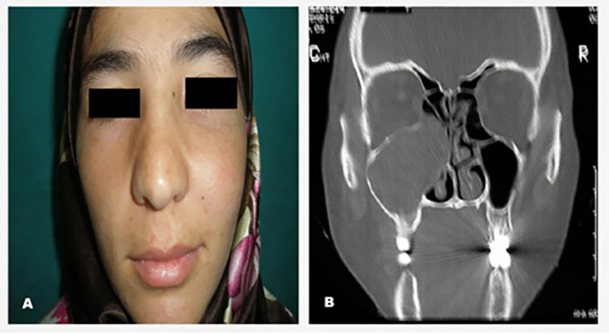
Figure 7. In the photograph, expansion of the maxillary bone and in CT scan; medial expansion of the wall of the maxillary sinus is seen.
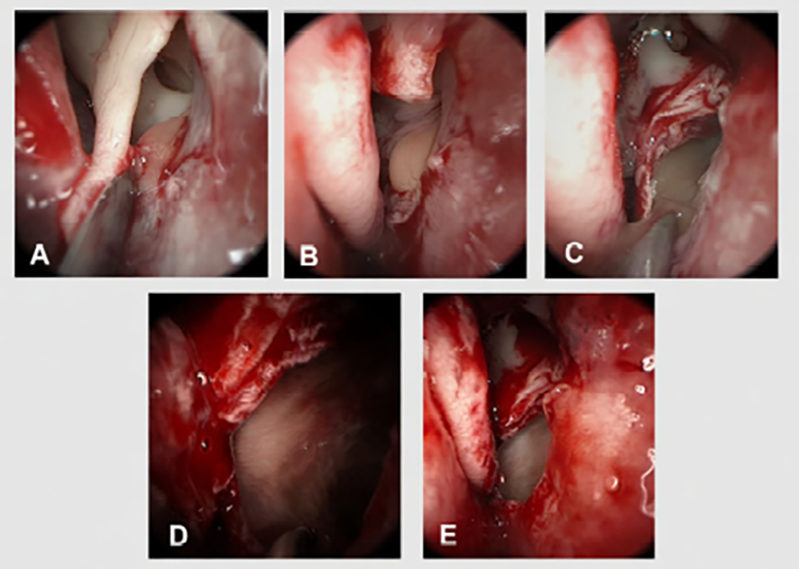
Figure 8. Endoscopic marsupialization of the maxillary sinus mucocele.
Additional Information
Publisher’s Note
Bayrakol MP remains neutral with regard to jurisdictional and institutional claims.
Rights and Permissions

About This Article
How to Cite This Article
Özer Erdem Gür, Mustafa Kaymakcı, Nevreste Didem Sonbay Yılmaz. Paranasal sinus mucoceles. Eu Clin Anal Med 2016;4(3):90-95. doi:10.4328/ECAM.97
- Received:
- June 6, 2016
- Accepted:
- June 11, 2016
- Published Online:
- September 1, 2016
- Printed:
- September 1, 2016