
Abstract
AimDiffering information about the muscularis mucosa layer of the esophagus can be found in reference books. We aimed to reevaluate the organization of the muscularis mucosa layer based on the examination of sections of esophagi obtained from cadavers.MethodsTen cadaver specimens were used in our study. Tissue sections were obtained from the level of the cricoid cartilage and of esophageal regions in close proximity with the gastroesophageal junction. These slices underwent routine histological tissue procedures and were embedded into paraffin blocks. They were stained with Hematoxylin-Eosin and Masson’s Trichrome and examined under a light microscope.ResultsWe observed that the muscularis mucosa layer exists on esophagus sections obtained from the cricoid level. When muscularis mucosa layers of proximal and distal esophageal regions were compared, it was noted that the layer of the distal region was thicker than the proximal one.ConclusionThere are muscularis mucosa layers in regions located at both the cricoid cartilage level and near the gastroesophageal junction regions; the layer in the proximal segment is thinner.
Keywords
Introduction
The esophagus is a muscular tube 25 cm long, located between the pharynx and the stomach. It allows for the transmission of food to the stomach. It begins in the cricoid cartilage, at the sixth cervical vertebrae. It passes through the diaphragm, at the tenth thoracic vertebrae level. It terminates level with the eleventh thoracic vertebrae at the gastric cardia. The esophagus consists of three parts, the cervicalis, toracalis, and the abdominalis.1 The wall of the esophagus consists of four layers. From the lumen outward, they are as follows: mucosa, submucosa, muscularis externa, and adventitia.
There is differing information related to the arrangement of the muscularis mucosa, a muscular layer located on the medial side. A number of anatomy and histology reference books have written that there is no muscularis mucosa layer in the proximal segment of the esophagus or that it is very thin1-2 Other books have written that this layer exists3-4 on the proximal segment. Moreover, there is also differing information about the distribution of the muscularis mucosa layer in the proximal and distal segments. There is some information indicating that it is a thin layer, interrupted in the proximal segment and uninterrupted in the distal segment.5 and that muscularis mucosa gets thicker from the proximal to the distal segment.1,4
The aim of our study was to compare information from different reference books and to examine human cadaver esophageal specimens to reevaluate this information. We believe that clinicians and surgeons will benefit from the results of our study
Materials and Methods
The study was performed with the approval of the local ethics committee, (2016-04, 05/5). It was carried out with samples taken from 10 (6 male/4 female) fixed cadavers. The age range of the cadavers was 55-70 years of age, with a mean age of 62. None of the cadavers had a known medical history of hiatal hernia or abdominal or thoracic surgery. Following the opening of the thoracic and abdominal cavities, the diaphragm was well dissected away from the anterior abdominal wall.
Next, the liver, heart, and lungs were retracted or dissected away to expose the esophagopharyngeal-gastroesophageal region. The esophagus was cut at the cricoid cartilage level, between vertebra thoracalis 10-11. Cadavers samples first underwent gross examination (Figure 1). Cadaver tissue specimens were fixed in formalin for light microscopic analyses. After fixation, the esophagus tissues were processed routinely for embedding in paraffin. Tissue sections of 5 µm were stained with Hematoxylin-Eosin (H&E) and Masson’s Trichrome to evaluate the muscularis mucosa layer of the esophagus. The sections were examined under a light microscope.
Results
A total of ten esophagus sections of cadavers were examined. Muscularis mucosa layers were found on the sections obtained from the cricoid cartilage level (Figure 2A-2B). We observed that the muscularis mucosa layer on the sections obtained from the cricoid level were thinner than those obtained from the esophagogastric level (Figure 3A, B, C and D).
Discussion
The esophagus transports food from the pharynx to the stomach. In the thorax and abdominal cavity, the esophagus is located in front of the spine. It passes into the posterior mediastinum from the superior mediastinum.6 The esophagus is made up of four concentric layers: tunica mucosa, tunica submucosa, tunica muscularis externa, and tunica adventitia. The mucosa is the innermost layer of the esophagus. It consists of a covering of nonkeratinized stratified squamous epithelium. The underlying lamina propia is loose connective tissue. The deep layer of smooth muscle is called the muscularis mucosa. Muscularis externa is the upper one-third, composed of striated muscle. Striated muscle and smooth muscle fibers are found in the middle third of the esophagus. The lower third of the esophagus is composed entirely of smooth muscle.3-4 This distribution of muscle fibers ensures that the swallowed pieces of food pass quickly through the upper part and slowly through the lower part of the esophagus.1 There are differences related to the arrangement of the muscularis mucosa layer among reference books.1-2-3 In the Introduction of a Cell Histology and Pathology book, it is written that there is no muscularis mucosa layer in the proximal esophagus and it is seen in the region adjacent to the stomach.2 Similarly, in Gray’s Anatomy it is mentioned that there is no muscularis mucosa layer in the proximal esophagus or it is very thin.1 Another study indicated that the muscularis mucosa layer in the esophagus starts at the proximal level and consists of longitudinal smooth muscles.3,7
In our study, we observed that there is a muscularis mucosa layer on sections obtained from the cricoid cartilage level. Little is known about the physiologic role of the muscularis mucosa during peristalsis and it is assumed that it provides for the movement of mucosa.7 On the other hand, it is considered that muscularis mucosa in the proximal esophagus has the function of aiding swallowing.3,5 In both Gray’s Anatomy and Human Histology, it is stated that the muscularis mucosa layer has been observed as thickening from the prox imal segment down to the distal segment.1,4 We observed that the muscularis mucosa layer on sections obtained from the cricoid cartilage level are thinner in comparison with those from the esophagogastric level. Some studies have reported that the muscularis mucosa layer of the esophagus gets thicker from the proximal segment to the distal segment, in agreement with our own study.7-8
Declarations
Informed Consent
Informed consent was obtained from all participants.
Conflict of Interest
The authors declare no conflicts of interest.
Funding
None.
References
- Standring S, Gray H. Gray’s anatomy. 40th ed. New York, NY: Churchill Livingstone; 2008:949-952.
- Kierszenbaum AL, Tres LL. Histology and cell biology: an introduction to pathology. 4th ed. Philadelphia, PA: Elsevier; 2016:482.
- Ross MH, Pawlina W. Histology: a text and atlas. 6th ed. Philadelphia, PA: Lippincott Williams & Wilkins; 2011:572.
- Lowe JS, Anderson PG. Stevens & Lowe’s human histology. 4th ed. Philadelphia, PA: Elsevier; 2015:199.
- Eşrefoğlu M. Özel histoloji. 2. baskı. İstanbul: İstanbul Medikal Yayıncılık; 2016:104.
- Sinnatamby CS. Last’s anatomy: regional and applied. 10th ed. New York, NY: Churchill Livingstone; 1999:201.
- Paterson WG. Esophageal peristalsis. GI Motility Online. 2006.
- Christensen J, Wingate DL, Gregory RA. A guide to gastrointestinal motility. Oxford: Butterworth-Heinemann; 1983.
Figures
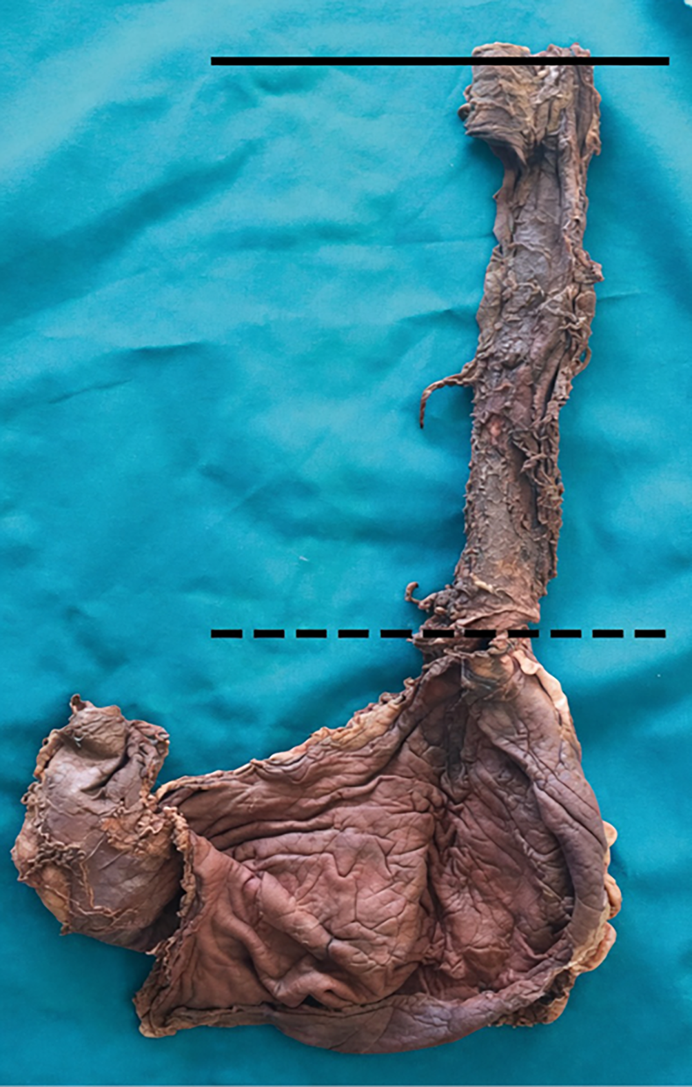
Figure 1. The solid line is level with the esophagopharyngeal junction. The dashed line is level with the gastroesophageal region.

Figure 2. Cartilage cricoid level of esophagus; (A) muscularis mucosa (black arrow), lamina propia (black line), and muscularis externa (black star) (Masson’s Trichrome staining, Scale bar 200 µm). (B) nonkeratinized stratified squamous epithelium (yellow star), lamina propia (yellow line), and muscularis mucosa (yellow arrow) (Masson’s Trichrome staining, Scale bar 100 µm)
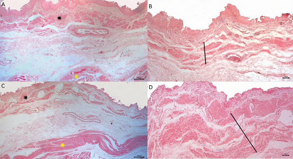
Figure 3. Cartilage cricoid level of esophagus; (A) muscularis mucosa (black star), and muscularis externa (yellow star) (H&E, Scale bar 200 µm). (B) muscularis mucosa (black line) (H&E, Scale bar 50 µm). Gastroesophageal region of esophagus; (C) muscularis mucosa (black star) and muscularis externa (yellow star) (H&E, Scale bar 200 µm). (D) muscularis mucosa (black line) (H&E, Scale bar 50 µm).
Additional Information
Publisher’s Note
Bayrakol MP remains neutral with regard to jurisdictional and institutional claims.
Rights and Permissions

This work is licensed under a Creative Commons Attribution-NonCommercial 4.0 International License (CC BY-NC 4.0). To view a copy of the license, visit https://creativecommons.org/licenses/by-nc/4.0/
About This Article
How to Cite This Article
Esra Erdoğan, Nurcan Ercıktı, Yasin Ilgaz. Mucosa muscularis of esophagus; revisiting of on the adaver. Eu Clin Anal Med 2026;4(3):87-89. doi:10.4328/ECAM.98
- Received:
- 06.06.2016
- Accepted:
- 10.06.2016
- Published Online:
- 01.09.2016
- Printed:
- 01.09.2016