
Abstract
IntroductionThe plantar area is different from other parts of the skin because its unique structure is resistant to external stress from physical activities. However, hyperkeratotic local skin thickening due to standing for long periods of time, recurrent minor traumas, unsuitable shoe choice, or bone deformity is called a callus.Case PresentationThis condition, which is seen in more than 78% of the population, is more frequent in older populations. Calluses may cause workload loss, difficulty in carrying out daily activities, and falls by causing pain and difficulty in walking. There are various methods of treatment and these methods vary depending on the characteristics of the condition.ConclusionThis study is the case report of a 67-year-old female patient who had multiple calluses on one foot.
Keywords
Introduction
Calluses are frequent problems, affecting the feet of a great majority of the population, especially the elderly. Large series of studies have reported one or more calluses in people over the age of 65 with a rate of 58.2%.1 Calluses are the third most common deformity of the feet after nail disorders and lesser toe problems.1 Calluses are common in older populations, particularly on the plantar; they are 4 times more common in women than in men.2 Calluses are considered to be a reaction to prolonged standing and extreme mechanical stresses.3 Most common plantar calluses are under the third and fourth metatarsal heads. Dorsal and interdigital calluses are less common than plantar calluses.3 Multiple calluses on the hands and feet are seen in diseases such as palmoplantar hyperkeratosis and congenital paronychia that are related to skin.4 However, isolated multiple calluses on one foot have not been reported. This study is a case report of multiple calluses on one foot, which is rare in the literature.
Discussion
Plantar area skin is different from other parts of the skin because of its unique structure.5 The part which contains keratin, the stratum corneum, protects the skin against external stress and physical activity. The stratum corneum is not a dead layer but it provides natural moisturizing factors, such as water and oil, and other protective adaptive functions. If dryness occurs in the stratum corneum as the result of a deterioration in the water balance, corneodesmosomes gradually degrade and then peeling and chapping occur in the corneocytes. Hypertrophy which occurs in the stratum corneum as a result of recurrent traumas of the skin is called a hyperkeratotic callus. Calluses, which are seen in more than 78% of the population, are more common in older populations and women.6 A callus is a hyperkeratotic lesion which contains a keratin nucleus that causes pain and inflammation. This lesion develops as a result of an increase in keratinocyte activity caused by chronic pressure and skin eruption.6 The use of unsuitable shoes and bone deformities that cause abnormal pressure provoke these lesions. Two typical places for these lesions are the fifth toe dorsolateral side and the areas that are exposed to pressure on the foot plantar side. A chronic nucleus forms as a result of mechanical stress. It tries to protect the skin bycausing the growth of hyperkeratotic tissue. Increased chronic pressure causes the process to enter a vicious circle and eventually the tissue becomes rigid and whiter in a coniform shape, characterized by the presence of a nucleus surrounded by a yellowish circular area. This results in painful and rigid skin eruptions which then causeadditional dryness.7 The pain is caused by the pressure of the cone-shaped nucleus on the nerve endings of the dermis.8 This pain causes a restriction in mobility as it becomes difficult to perform daily activities. Falls occur as a result of the pain that does not allow the patient to step in a complete and balanced way. Our casewas 67 years old and had been doing farm work which caused continuous trauma. There are no reports of isolated multiple calluses of one foot in the literature, making our case report unique. Diabetes is closely related with hyperkeratotic lesions. Neuropathy in diabetes can cause unsuitable pressure points, eruptions, recurrent traumas, ulcers, and even dramatic outcomes that end in amputation.9 Our case had no comorbid diseases except hypertension. Verruca, a viral infectious disease frequently mistaken for callus, should be considered in definitive diagnosis. In verrucae, keratinocytes are infected by the human papilloma virus to form hyperkeratosis. The lesion head is in the form of an inward cone and its overgrowth presses with walking and standing, causing pain. Although there is generally a single verruca, multiple lesions can be seen in immunosuppressive patients. To distinguish the 2 lesions, in calluses, it is painful when stepping on the lesion, but in verruca, it is painful when the edges of the lesion are pressed together. In addition, the presence of a nucleus and the continuation of skin lines in the callus are distinctive.For the treatment of calluses, suitable shoe use, foot pads, adhesive tape, moisturizers, solutions that contain salicylic acid, bleomycin, keratolytic drugs, cryotherapy, cauterization, debridement, and total excision are precautions to prevent the formation of bone deformities and the need for further treatment.6,8,10 Our case, who had not yet received any treatment, was informed and sent to the dermatology department to develop a treatment plan.
Conclusion
Calluses are frequently on the plantar, especially in older populations and women. In jobs which require prolonged standing, such as farming, mechanical stress can cause serious skin problems. We believe that various precautions should be taken in the early term to prevent morbidity and to ensure foot health in groups with increased risk factors.
Declarations
Informed Consent
Written informed consent was obtained from the patient for publication of this case report and accompanying images.
Conflict of Interest
The authors declare no conflicts of interest.
Funding
None.
References
- Dunn JE, Link CL, Felson DT, Crincoli MG, Keysor JJ, McKinlay JB. Prevalence of foot and ankle conditions in a multiethnic community sample of older adults. Am J Epidemiol. 2004;159:491-498.
- Menz HB, ed. Foot problems in older people: assessment and management. Churchill Livingstone Elsevier; 2008.
- Yates B, ed. Merriman’s assessment of the lower limb. Churchill Livingstone Elsevier; 2009.
- Tejinder Kaur T, Rani R, Gupta RR, Mahajan BB, Sachdeva R. Pachyonychia congenita type 1: Jadassohn-Lewandowsky syndrome. Egypt Dermatol Online J. 2010;6(2):15.
- Swensson O, Langbein L, McMillan JR, Stevens HP, Leigh IM, McLean WH, et al. Specialized keratin expression pattern in human ridged skin as an adaptation to high physical stress. Br J Dermatol. 1998;139(5):767-775. doi:10.1046/j.1365-2133.1998.02499.x
- Hashmi F, Nester C, Wright C, Newton V, Lam S. Characterising the biophysical properties of normal and hyperkeratotic foot skin. J Foot Ankle Res. 2015;8:35. doi:10.1186/s13047-015-0092-7
- Hashmi F, Wright C, Nester C, Lam S. The reliability of non-invasive biophysical outcome measures for evaluating normal and hyperkeratotic foot skin. J Foot Ankle Res. 2015;8:28. doi:10.1186/s13047-015-0083-8
- Lee WJ, Lee SM, Won CH, Chang SE, Lee MW, Choi JH, et al. Efficacy of intralesional bleomycin for the treatment of plantar hard corns. Int J Dermatol. 2014;53(12):572-577. doi:10.1111/ijd.12586
- Tang UH, Zügner R, Lisovskaja V, Karlsson J, Hagberg K, Tranberg R. Foot deformities, function in the lower extremities, and plantar pressure in patients with diabetes at high risk to develop foot ulcers. Diabet Foot Ankle. 2015;6:27593. doi:10.3402/dfa.v6.27593
- Landorf KB, Morrow A, Spink MJ, Nash CL, Novak A, Potter J, et al. Effectiveness of scalpel debridement for painful plantar calluses in older people: a randomized trial. Trials. 2013;14:243. doi:10.1186/1745-6215-14-243
Figures
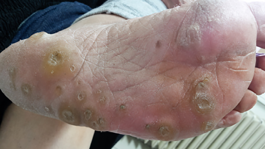
Figure 1. The appearance of multiple calluses on the left plantar, especially in areas which come into contact with water.
Additional Information
Publisher’s Note
Bayrakol MP remains neutral with regard to jurisdictional and institutional claims.
Rights and Permissions

This work is licensed under a Creative Commons Attribution-NonCommercial 4.0 International License (CC BY-NC 4.0). To view a copy of the license, visit https://creativecommons.org/licenses/by-nc/4.0/
About This Article
How to Cite This Article
Alper Çıraklı, Erdal Uzun, Yakup Ekinci, İsmail Büyükceran, Mustafa Deveci. Multiple calluses on the plantar surface of the foot: A Case Report. Eu Clin Anal Med 2026;4(2):65-67. doi:10.4328/ECAM.88
- Received:
- 19.04.2016
- Accepted:
- 04.05.2016
- Published Online:
- 04.05.2016
- Printed:
- 04.05.2016