Dilated cardiomyopathi mimicking massive pleural effusion in an infant
Cardiomyopathy mimicking pleural effusion
Authors
While pneumonia incidence is decreasing, pneumonic complications such as pleural effusion and empyema are increasing across the world. In the presence of pleural fluid, diagnostic thoracentesis is used to evaluate the composition of the fluid. Performing thoracentesis in the case of an uncertain diagnosis of pleural effusion may lead to life-threatening complications.1,2
A 6-month-old infant was diagnosed with right-side pneumonia and treated for respiratory failure in the pediatric intensive care unit. Thoracic surgery was consulted due to a massive pleural fluid appearance on a chest X-ray (Figure 1A). Thoracic ultrasonography performed before thoracentesis revealed no pleural fluid or consolidation, but rather cardiomegaly and compressed lung parenchyma in the left hemithorax. Consequently, no thoracentesis was performed. Echocardiography revealed "moderate to severe mitral valve insufficiency and tricuspid insufficiency with dilated cardiomyopathy and congestive heart failure." After five days of follow-up under mechanical ventilation and medical treatment for pneumonia and heart failure, the chest X-ray findings and clinical status dramatically improved (Figure 1B, C, D).
Thoracentesis is an invasive diagnostic procedure that is used frequently. In cases involving a potential misdiagnosis of pleural fluid accumulation, clinicians should not be too hasty in performing a thoracentesis. For patients who are not clinically stable enough for computed tomography, thoracic ultrasound is an effective imaging modality to guide the clinician.3,4,5
Declarations
Animal and Human Rights Statement
All procedures performed in this study were in accordance with the ethical standards of the institutional and/or national research committee and with the 1964 Helsinki Declaration and its later amendments.
Informed Consent
Informed consent was obtained from all participants.
Data Availability
The datasets used and/or analyzed during the current study are not publicly available due to patient privacy reasons but are available from the corresponding author on reasonable request.
Conflict of Interest
The authors declare no conflicts of interest.
Funding
None.
Scientific Responsibility Statement
The authors declare that they are responsible for the scientific content of the article, including the study design, data collection, analysis and interpretation, manuscript preparation, and approval of the final version of the manuscript.
References
-
Muzumdar H, Arens R. Pleural fluid. Pediatr Rev. 2007;28(12):462-464. doi:10.1542/pir.28-12-462
-
Rocha G. Pleural effusions in the neonate. Curr Opin Pulm Med. 2007;13(4):305-311. doi:10.1097/mcp.0b013e3281214459
-
Lichtenstein DA. Ultrasound examination of the lungs in the intensive care unit. Pediatr Crit Care Med. 2009;10(6):693-698. doi:10.1097/pcc.0b013e3181b7f637
-
Singhi SC, Mathew JL, Kumar RM, et al. Clinical pearls in pediatric cardiology. Indian J Pediatr. 2011;78(10):1273-1280. doi:10.1007/s12098-011-0466-1
-
Loizzi M, De Palma A, Pagliarulo V, Loizzi D, Sollitto F. Pulmonary infections of surgical interest in childhood. Thorac Surg Clin. 2012;22(3):387-401. doi:10.1016/j.thorsurg.2012.04.005
Figures
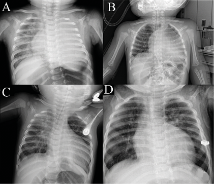
Figure 1. The opacity fullfilling left hemithorax, moving mediastinum to opposite site suggests a massive pleural effusion
(A). Second day chest x ray of the patient received medical treatment and mechanic ventilation (B). The fifth day of the treatment (C). The chest x ray of the extubated patient demonstrates waned cardiac size and expanded left lung (D).
Additional Information
Publisher’s Note
Bayrakol MP remains neutral with regard to jurisdictional and institutional claims.
Rights and Permissions

This work is licensed under a Creative Commons Attribution-NonCommercial 4.0 International License (CC BY-NC 4.0). To view a copy of the license, visit https://creativecommons.org/licenses/by-nc/4.0/
About This Article
How to Cite This Article
Naime Tokur, Sevki Demiroz, Burcu Cantay . Dilated Cardiomyopathi Mimicking Massive Pleural Effusion in an Infant. Eu Clin Anal Med 2014; DOI:10.4328/ECAM.25
- Received:
- December 19, 2013
- Accepted:
- December 28, 2013
- Published Online:
- January 1, 2014
- Printed:
- January 1, 2014