Dental extraction under deep sedation in a patient with rubinstein taybi syndrome: A Case Report
Deep sedation and rubinstein-taybi syndrome
Authors
Abstract
Introduction,Rubinstein-Taybi Syndrome (RTS), is a genetic disorder caused by a heterozygous mutation on chromosome 16. This multiple congenital anomaly syndrome is characterized with mental retardation, craniofacial deformities and finger anomalies. Children with RTS generally encounter severe dental problems and need interventions under sedation or general anesthesia.
Case PresentationA 14-yr old boy with RTS is scheduled for dental treatment at Gazi University Faculty of Dentistry. Preoperative physical findings of the patient with limited cooperation revealed microcephalia, retrognatia, and broad thumbs. His Mallampati score was II.
After placing an intravenous cannula and establishing standard monitarization, 50/50% O2/N2O was administered via nasal mask while maintaining spontaneous ventilation. Afterwards, midazolam and ketamine were given to obtain desired level of sedation.
ConclusionAlthough treatment under deep sedation has been performed without any adverse events for this patient, we believe that all the precautions mentioned in the algorithms should strictly be taken against possible difficult airway.
Keywords
Introduction
Rubinstein Taybi Syndrome (RTS) is a genetic disorder caused by heterozygous mutation on chromosome 16p13 and is characterized by short stature, moderate to severe intellectual disability, distinctive facial features, and broad thumbs and first toes. The prevalence is 1 in 100,000-125,000 births.1,2
Poor oral hygiene in RTS patients results in dental problems. Limited cooperation with these patients negatively affects the dental treatment protocols. Therefore, general anesthesia or sedation is required for the patients with RTS for dental treatment.
Specific problems related to the disease such as craniofacial deformities, cardiac anomalies and high incidence of gastroesophageal reflux (GER) raise the risks related to anesthesia.1 There is limited data on the literature about the deep sedation practice in patients with RTS. We aimed to describe our experience of deep sedation for dental treatment in a child with RTS.
Case Presentation
A 14-yrs old boy with RTS is scheduled for dental treatment at Gazi University, Faculty of Dentistry. He had two prior operations under general anaesthesia for correction of patent ductus arteriosis and undescended testis. Preoperative physical findings revealed craniofacial anomalies including microcephalia, retrognatia, and broad thumb. He had also kyphoscoliosis. A slight loss of strength in the extremities was also observed. His Mallampati score was II and laboratory findings were within normal limits.
The patient was taken to treatment unit without any premedication and Lactated Ringer infusion was started. Noninvasive blood pressure, peripheral oxygen saturation, ECG and ETCO2 were monitored and 50/50% N2O/O2 was administered through a nasal mask (Figure 1).
While maintaining spontaneous ventilation, 0.03 mg/kg midazolam and 1 mg/kg ketamine were given intravenously. The Ramsay sedation score of the patient was 5 without further medication during the whole procedure which lasted 15 minutes. Suction was placed in the patient’s mouth for aspirating the oral contents during the procedure. Number 36 and 46 impacted molars were extracted. The patient was transferred to postanaesthesia care unit and discharged after 4 hours of close monitoring.
Discussion
Patients with RTS has typical facial morphological abnormalities and clinical features such as hypoplastic maxilla, highly arched palate, tonsillar hypertrophy, adenoid formation, feeding intolerance, hypotonia, GER, motor and mental retardation which have potential to increase the morbidity during anaesthesia and sedation procedures.1 Typical facial expression (Figure 1), retrognatia and mental retardation of the patient were the potential problems for the planned sedation procedure in our patient.
Dental problems due to poor oral hygiene are observed in 67% of the patients with RTS.2 Repeated dental examinations or treatments are usually impossible to perform under local anaesthesia because of limited cooperation. There are few reports on the general anaesthesia practice of the patients with RTS in the English literature but we were unable to find much data about the deep/unconscious sedation practice.1,2
A paedodontist, Morales-Chaves2 also reported a 13-yrs old girl who had dental treatment for dentoalveolar abscess and crowded teeth under conscious sedation. During sedation procedure with midazolam and ketamine, he experienced respiratory difficulties treated by an anaesthesiologist on duty without any adverse effect. There is limited data about the anaesthetic agents that could be used in patients with RTS.
Neostigmine and atropine are the agents that should be avoided as they can trigger the ectopic rhythms due to the changes in sympathetic and parasympathetic activity. Succinylcholine may cause supraventricular tachycardia, premature atrial and ventricular contractions. Therefore it is also not recommended.3,4 As the patient had the history of a corrective heart surgery, we preferred deep sedation of which we could avoid the anaesthetic agents that has the potential of provoking ectopic rhythms.
Patients with RTS are candidates for difficult intubation due to craniofacial anomalies they have.3 Aspiration pneumonia (AP) risk related to GER and history of recurrent respiratory tract infections should also be considered during interventions for RTS. However, Twigg and Cook5 reported an uneventful general anaesthesia practise in RTS patient which they placed Proseal laryngeal mask for airway control. They thought that laryngeal mask will not increase the risk of AP as the patient had no history of GER.
The choice of deep sedation may be criticized for the risk of AP in our patient but as in the previous literature our patient had no history and findings of AP such as recurrent pulmonary infections. The head, neck and temporo-mandibular joint movements of the patient were within normal limits. Also the Mallampati score was II. Therefore no difficulties were anticipated for maintaining the patency of the airway but all the precautions were taken according to the difficult airway algorithm. Also, suction was placed in patient’s mouth to protect aspiration of the oral contents.
The patient’s planned dental treatment was completed under deep sedation without any adverse event. As a result, we believe that deep sedation can also be performed safely by an anaesthesia team experienced in paediatric anaesthesia and in the presence of decent equipment, for dental treatments of patients with RTS.
Why this paper is important?* The choice of anaesthesia is important for postoperative quality of life of the disabled and children.
* General anaesthesia has some disadvantages such as hemodynamic instability especially in patients with cardiac problems as in RTS.
* Deep sedation might be a appropriate choice of anaesthesia procedure in selected group of disabled patients.
Declarations
Animal and Human Rights Statement
All procedures performed in this study were in accordance with the ethical standards of the institutional and/or national research committee and with the 1964 Helsinki Declaration and its later amendments.
Informed Consent
Written informed consent was obtained from the patient for publication of this case report and accompanying images.
Conflict of Interest
The authors declare no conflicts of interest.
Funding
None.
Scientific Responsibility Statement
The authors declare that they are responsible for the scientific content of the article, including the study design, data collection, analysis and interpretation, manuscript preparation, and approval of the final version of the manuscript.
References
-
Wiley S, Swayne S, Rubinstein JH, Lanphear NE, Stevens CA. Rubinstein-Taybi syndrome medical guidelines. Am J Med Genet A. 2003;119A:101-110. doi:10.1002/ajmg.a.10009
-
Morales-Chavez MC. Dental management of a patient with Rubinstein-Taybi syndrome. Spec Care Dentist. 2010;30:124-126.
-
Stirt JA. Anesthetic problems in Rubinstein-Taybi syndrome. Anesth Analg. 1981;60:534-536. doi:10.1213/00000539-198107000-00016
-
Stirt JA. Succinylcholine in Rubinstein-Taybi syndrome. Anesthesiology. 1982;57:429. doi:10.1097/00000542-198211000-00025
-
Twigg SJ, Cook TM. Anaesthesia in an adult with Rubinstein-Taybi syndrome using the ProSeal laryngeal mask airway. Br J Anaesth. 2002;89:786-787. doi:10.1093/bja/aef248
Figures
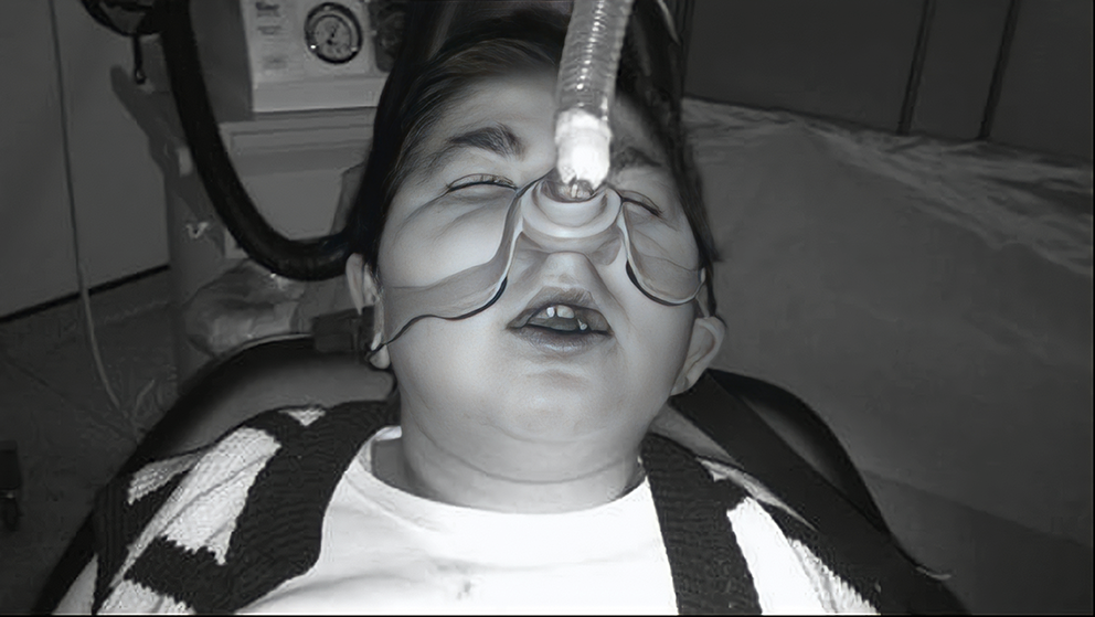
Figure 1. Administration of 50/50 % N2O/O2 through a nasal mask while maintaining spontaneous ventilation
Additional Information
Publisher’s Note
Bayrakol MP remains neutral with regard to jurisdictional and institutional claims.
Rights and Permissions

This work is licensed under a Creative Commons Attribution-NonCommercial 4.0 International License (CC BY-NC 4.0). To view a copy of the license, visit https://creativecommons.org/licenses/by-nc/4.0/
About This Article
How to Cite This Article
Metin Alkan, Hasan Kutluk Pampal, Hakan Dayanir, Mustafa Arslan, Berrin Isik. Dental extraction under deep sedation in a patient with rubinstein taybi syndrome: A Case Report. Eu Clin Anal Med 2013;1(2):38-40. doi:10.4328/ECAM.10
- Received:
- April 24, 2013
- Accepted:
- May 9, 2013
- Published Online:
- May 9, 2013
- Printed:
- May 9, 2013