In silico study of Ferula latisecta-derived compounds molecular interactions with α-glucosidase
Molecular interactions of Ferula latisecta
Authors
Abstract
AimOur study aimed to investigate the sulfur compounds' antidiabetic effect in Ferula latisecta.
MethodsThe molecular docking method investigated the α-glucosidase inhibitory effect of the components. In our study, the pharmacokinetic properties of Ferula latisecta compounds were also investigated with the SwissADME method, and the toxicity risk analyzes were investigated with Protox II tools. Ferula latisecta compounds were drawn from the literature in Chemdraw and and α-glucosidase enzyme structure was obtained from Protein Data Bank. Finally, the molecular interaction analysis between α-glucosidase and compounds from Ferula latisecta was performed by AutoDock 1.5.7. Molecular interactions were investigated using Discovery Studio Visualizer and Ligplot 2.1 program.
ResultsAll the selected sulfur compounds from Ferula latisecta followed Lipinski’s rules, had sufficient binding energy, and lacked toxicity; therefore, they were appropriate candidates for α-glucosidase inhibition. Among these compounds, 2-(4-hydroxyphenyl) ethyl lignocerate and isosco-poletin showed the lowest binding energy and the highest inhibitory effect on α-glucosidase enzyme with −9.1 and −7.7 kcal/mol, respectively.
ConclusionThese compounds also indicated a lower binding energy than the standard inhibitor (miglitol). Among the sulfur compounds in Ferula latisecta 2-(4-hydroxyphenyl) ethyl lignocerate and isoscopoletin were predicted to be the potent inhibitors due to having more hydrogen bonds and hydrophobic interactions with the active site of α-glucosidase.
Keywords
Introduction
Diabetes mellitus (DM) continues to be a significant threat to human health today, and it is estimated that by 2045, the number of patients worldwide will exceed 642 million.1 DM is a chronic metabolic disease characterized by high blood sugar levels due to insulin resistance.1,2 High blood glucose levels also damage blood vessels and nerves, causing various health problems such as hypertension, cardiovascular disease, blindness, stroke, amputations, kidney, and dental conditions.3,4,5 Although different drug treatments are available today, the side effects of drugs continue to be a problem.
Antidiabetic drugs fall into several categories, including sulfonylureas, bioguanidines, insulin mimetics (glucagon-like peptide analogs), and α-glucosidase inhibitors.
The discovery of new drugs is essential because of the side effects of existing antidiabetic drugs. Academic studies have recently confirmed medicinal plants' beneficial effects in treating diabetes.6,7 Researched impacts of plants include improving glycemic control, lowering serum lipid levels, inhibiting oxidative stress, and improving inflammatory response.8,9
The genus Ferula is mainly distributed throughout central and Southwest Asia (especially Iran and Afghanistan), the Far East, North India, and the Mediterranean. The main phytochemicals present in the genus are as follows: coumarin, coumarin esters, sesquiterpenes, sesquiterpene lactones, monoterpene, monoterpene coumarins, prenylated coumarins, sulfur-containing compounds, phytoestrogen, flavonoids, and carbohydrates.10
People used these plants' roots, leaves and fruits as vegetables, spices, or medicine. In addition, other species of this genus have been reported to be popularly used for sedatives, digestive disorders, rheumatism and arthritis, neurological disorders, inflammations, dysentery, headaches, and toothaches.11,12
Ferula latisecta (F. latisecta) has been used in Iranian folk medicine to treat parasitic diseases, relieve stomachaches in infants, and to control diabetes. In other studies, it has been shown that essential oil obtained from the above-ground parts of F. latisecta has an antimicrobial effect.13,14
In a previous study, streptozotocin-induced diabetic male Wistar rats were treated with F. latisecta root (400 mg/kg/day) for four weeks, resulting in significantly reduced LDL levels in the kidney and liver (p < 0.05) and thiol (p < 0.05) and superoxide dismutase (p < 0.01). In addition, the root of F. latisecta was found to reduce serum total cholesterol levels (p < 0.05) and prevent the progression of hyperglycemia.15
The mechanism of action of many drugs used to treat diabetes occurs through the inhibitory effect of α-glucosidase (AG). AG is a carbohydrate hydrolyzing enzyme. Overactivity of α-glucosidase causes glucose to be absorbed by the small intestinal lumen and enter the bloodstream. Glucose release results in increased blood glucose levels and uncontrolled hyperglycemia in type-2 DM patients. α-Glucosidase is one of the glucosidase enzymes responsible for the mechanism of DM. α-glucosidase catalyzes the hydrolysis reaction by breaking the α-(1,4)-glycosidic bond of carbohydrates to become a free monosaccharide (α-D-glucose) before entering the bloodstream. The AG enzyme plays a vital role in carbohydrate metabolism by converting starch and disaccharides into glucose.16 As a result of inhibiting the enzyme α-glucosidase, carbohydrate digestion slows down and causes glucose to enter the bloodstream later. As a result, it constitutes a proper prophylactic treatment for hyperglycemia.17 Therefore, inhibition of AG is a practical therapeutic approach for treating DM.
Our study aims to determine the toxicity of eight compounds12 of F. latisecta by in silico methods and investigate the anti-diabetes effect by examining the inhibitory effect of the compounds on the α-glucosidase enzyme by molecular docking method.
Materials and Methods
First, important sulfur-containing phenolic compounds were obtained from the literature on F. latisecta.12 Next, compounds were drawn using ChemDraw software. Finally, a suitable crystallographic structure with a resolution of 1.6oA with the code PDB ID 3A4A from the α-glucosidase enzyme protein data bank was obtained from the protein database.
Determination of Pharmacological Properties of Compounds by Lipinski ParameterThe potentially effective sulfur compounds of F. latisecta were evaluated using the Lipinski parameter to inhibit the activity of the α-glucosidase enzyme. All the compounds showed the properties required by Lipinski’s parameter; therefore, they were predicted to have optimal adsorption. The SwissADME database was used to obtain the Lipinski properties of the compounds.
Evaluating the Toxicity of the Selected CompoundsF. latisecta contains many sulfur compounds. Among the most highlighted sulfur compounds, latisulfide A, latisulfide B, latisulfide C, latisufide D, and latisulfide E, τ-cadinol, 2-(4-hydroxyphenyl) ethyl lignocerate and isoscopoletin are also found in this plant. Phenolic compounds: Lack of toxicity is one of the critical factors for choosing a compound as a therapeutic candidate. Therefore, in this study, the toxicity of each of the sulfur compounds, such as liver toxicity, carcinogenicity, immunotoxicity, mutagenicity and the toxicity class of the compounds were examined using the Protox tool.18 In addition, the accuracy of the toxicity analysis performed with the PKCSM software was checked.
Molecular Interaction AnalysisThis study used the Autodock vina tool (Version 1.1.4)19 to investigate the molecular interaction between α-glucosidase enzyme and selected ligands. Before the docking analysis, the enzyme structure was optimized by removing excess ligands and water molecules using the BIOVIA Discovery Studio 2021 program. The Spartan 14 (Version 1.1.4) program optimizes all compounds for energy. Kollman charges were determined by adding polar hydrogens to the protein using the AutoDock vina 1.5.7 tool. The partial load of the compounds was calculated using Compute Gasteiger in AutoDock 1.5.7 tool.
The x, y, and z coordinates were determined to bind the α-glucosidase enzyme to its catalytic site (x:20.53, y: -10.11 z:22.38 x:40, y:40 z:40). Finally, molecular interactions and binding types between the selected compounds and the α-glucosidase enzyme were investigated using the Discovery Studio visualizer and Ligplot (version 2.2.8) programs.20
Ethical ApprovalNot required for this in silico study.
Results
Pharmacological Properties of the Selected Compounds The selected ligands: latisulfide A, latisulfide B, latisulfide C, latisufide D, latisulfide E, τ-cardinal, 2-(4-hydroxyphenyl) ethyl lignoceric and isoscopoletin were obtained from the literature and drawn in ChemDraw software.12
Toxicity Analysis of the Selected CompoundsThe compounds were also evaluated for their toxicity and toxicity class. The criterion for selecting a compound as a drug candidate is the safety of the compound, and the compound should not show any toxicity.
Compounds contained in F. latisecta were evaluated in terms of toxicity and toxicity class (Table 1). In the ADME study conducted on SwissADME software, it was found to be appropriate among the compounds in terms of pharmacokinetic properties, except for 2-(4-hydroxyphenyl) ethyl lignocerate. Toxicity estimation was evaluated with the protox II tool program. According to the protox II tool prediction program, it was observed that hepatotoxic and immunotoxic effects may occur in all compounds. However, when we look at the probability values, it has been determined that the risk of toxicity is low. Toxicity class 4 indicates low toxicity of the ingredients. When the PKCSM program, which is a different software, was examined separately, no toxicity was detected. Compounds contained in F. latisecta were evaluated in terms of toxicity and toxicity class (Table 1).
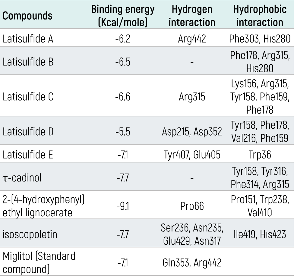
Molecular Interaction AnalysisThe results of the molecular interaction between the selected compound and the α-glucosidase enzyme obtained by molecular coupling assay are given in Table 2. All compounds showed good binding with the enzyme. The binding energy, hydrogen and hydrophobic interactions between the compounds and the enzyme are also presented in Table 3.
The results of the molecular docking study showed that all sulfur compounds in F. latisecta can bind to the active site of α-glucosidase and inhibit the activity of this enzyme. According to the docking analysis, the binding energies of the studied compounds are different; therefore, the critical energy ranges from –5.5 to –9.1 kcal/mol. The lower the binding energy level (negative), the stronger the binding between the receptor (enzyme) and ligands (compound or inhibitor). Among the selected sulfur compounds, 2-(4-hydroxyphenyl) ethyl lignoceric with a value of -9.1 kcal/mol and isoscopoletin with a weight of -7.7 kcal/mol showed the lowest binding energy. Therefore, they are estimated to provide the highest inhibitory effect among the compounds (Figure 1).
Previous studies have shown that amino acids such as LYS-156, GLN-279, HIS-280, PRO-312 GLU-277, ASP-352, ASN-415, and ARG-442 play a key role in the interaction of enzyme and inhibitor in the active site of α-glucosidase.21 Miglitol was used as a standard α-glucosidase inhibitor. The insertion results showed that miglitol, with a binding energy of -7.1 kcal/mol, has a significant binding energy and hydrogen bonding with amino acids Gln353 and Arg442.
Pro66 (3.60 and 5.50 Å) 2-(4-hydroxyphenyl) ethyl lignoceric complex and isoscopoletin complex with amino acids Ser236, Asn235, Glu429, Asn317 also have hydrogen bonds (Figure 2 a,b). Isoscopoletin showed more hydrogen bond interactions than other compounds. These two compounds have lower binding energies than other compounds. This also shows that it can bind more strongly to the enzyme. Figure 2 shows the three-dimensional view of the ionization, hydrophobic, polar, and hydrogen bonding reactions between α-glucosidase and isoscopoletin. The surface interactions of isoscopoletin within the active site of α-glucosidase are illustrated in Figure 3.
Discussion
In the present study, 2-(4-hydroxyphenyl) ethyl lignoceric has shown that F. latisecta has the lowest binding energy among the phenolic compounds. In another study, while the synthesis and inhibitory potentials of sulfur compounds were investigated, it was found that compounds such as methazolamide, acetazolamide, and timolol (having sulfur atoms with binding energies of -4.3, -4.8 and 4.5, kcal/mol in their structure, respectively) inhibited the α-glucosidase enzyme significantly.22 In this study, with a -9.1 kcal/mol value, 2-(4-hydroxyphenyl) ethyl lignocerate showed the best inhibition value among sulfur compounds.
In addition, miglitol did not show any hydrophobic interaction with α-glucosidase. However, it showed hydrophobic interactions with 2-(4-hydroxyphenyl) ethyl lignoceric and α-glucosidase Pro151, Trp238, and Val410. Exchange of isoscopoletin with α-glucosidase showed hydrophobic interaction with amino acids Ile419 and His423.
Conclusion
When we looked at the results of our study, it was clear that the pharmacokinetic properties of important sulfur compounds were good. Still, hepatotoxicity and immunotoxicity risk were observed in all components; however, the low toxicity probability levels and the toxicity class 4 show that it does not carry a significant stake in toxicity. Also, the hepatotoxic effects of hepatotoxic compounds were examined in a different software PKCSM program. When the data obtained from this program were analyzed, it was seen that they were not toxic. Based on the results of the docking study, it can be concluded that of the eight compounds in F. latisecta, 2-(4-hydroxyphenyl) ethyl lignocerate and isoscopoletin are more effective in inhibiting the α-glucosidase enzyme. These compounds also exhibited lower binding energies compared to miglitol. Therefore, they are likely to be more potent inhibitors than miglitol. Considering that in silico studies are used for preliminary studies and predictions, it is thought that the results of our study should be validated with in vivo and in vitro studies.
A comparison of 2-(4-hydroxyphenyl) ethyl lignocerate and isoscopoletin with miglitol is shown. Both selected compounds appear to interact with different amino acids in the catalytic pocket of the α-glucosidase enzyme.
Declarations
Animal and Human Rights Statement
This study did not involve human participants or animals.
Informed Consent
Not applicable.
Data Availability
The data generated or analyzed during this study are available from the corresponding author on reasonable request.
Conflict of Interest
The author declares no conflict of interest.
Funding
None.
Scientific Responsibility Statement
The authors declare that they are responsible for the article’s scientific content including study design, data collection, analysis and interpretation, writing, some of the main line, or all of the preparation and scientific review of the contents and approval of the final version of the article.
Abbreviations
AG: Alpha-glucosidase
ADME: Absorption, distribution, metabolism, and excretion
DM: Diabetes mellitus
LDL: Low-density lipoprotein
PDB: Protein Data Bank
References
-
Cho NH, Shaw JE, Karuranga S, et al. IDF Diabetes Atlas: global estimates of diabetes prevalence for 2017 and projections for 2045. Diabetes Res Clin Pract. 2018;138:271-281. doi:10.1016/j.diabres.2018.02.023
-
American Diabetes Association. 7. Diabetes technology: standards of medical care in diabetes—2021. Diabetes Care. 2021;44(suppl 1). doi:10.2337/dc21-s007
-
Cui Y, Zhang L, Zhang M, et al. Prevalence and causes of low vision and blindness in a Chinese population with type 2 diabetes: the Dongguan Eye Study. Sci Rep. 2017;7(1):11195. doi:10.1038/s41598-017-11365-z
-
Helgason CM. Blood glucose and stroke. Curr Treat Options Cardiovasc Med. 2012;14(3):284-287. doi:10.1007/s11936-012-0178-5
-
Zhang P, Gregg E. Global economic burden of diabetes and its implications. Lancet Diabetes Endocrinol. 2017;5(6):404-405. doi:10.1016/s2213-8587(17)30100-6
-
Ghorbani A. Best herbs for managing diabetes: a review of clinical studies. Braz J Pharm Sci. 2013;49(3):413-422. doi:10.1590/s1984-82502013000300003
-
Hosseini A, Shafiee-Nick R, Ghorbani A. Pancreatic beta cell protection/regeneration with phytotherapy. Braz J Pharm Sci. 2015;51(1):1-16. doi:10.1590/s1984-82502015000100001
-
Ghorbani A. Mechanisms of antidiabetic effects of flavonoid rutin. Biomed Pharmacother. 2017;96:305-312. doi:10.1016/j.biopha.2017.10.001
-
Prabhakar PK, Doble M. Mechanism of action of natural products used in the treatment of diabetes mellitus. Chin J Integr Med. 2011;17(8):563-574. doi:10.1007/s11655-011-0810-3
-
Sonigra P, Meena M. Metabolic profile, bioactivities, and variations in the chemical constituents of essential oils of the Ferula genus (Apiaceae). Front Pharmacol. 2021;11:608649. doi:10.3389/fphar.2020.608649
-
Dehghan G, Shafiee A, Ghahremani MH, Ardestani SK, Abdollahi M. Antioxidant potential of various extracts from Ferula szovitsiana in relation to their phenolic content. Pharm Biol. 2007;45(9):691-699. doi:10.1080/13880200701575098
-
Soltani S, Amin GR, Salehi-Sourmaghi MH, Schneider B, Lorenz S, Iranshahi M. Sulfur-containing compounds from the roots of Ferula latisecta and their cytotoxic activities. Fitoterapia. 2018;124:108-112. doi:10.1016/j.fitote.2017.10.012
-
Habibi Z, Salehi P, Yousefi M, et al. Chemical composition and antimicrobial activity of the essential oils of Ferula latisecta and Mozaffariania insignis from Iran. Chem Nat Compd. 2006;42(6):689-692. doi:10.1007/s10600-006-0253-9
-
Iranshahi M, Hassanzadeh-Khayat M, Bazzaz BSF, Sabeti Z, Enayati F. High content of polysulphides in the volatile oil of Ferula latisecta Rech. f. et Aell. fruits and antimicrobial activity of the oil. J Essent Oil Res. 2008;20(2):183-185. doi:10.1080/10412905.2008.9699986
-
Javanshir S, Soukhtanloo M, Jalili-Nik M, Yazdi AJ, Amiri MS, Ghorbani A. Evaluation potential antidiabetic effects of Ferula latisecta in streptozotocin-induced diabetic rats. J Pharmacopuncture. 2020;23(3):158-164. doi:10.3831/kpi.2020.23.3.158
-
Abdjan MI, Aminah NS, Kristanti AN, et al. Structure-based approach: molecular insight of pyranocumarins against α-glucosidase through computational studies. RSC Adv. 2023;13(6):3438-3447. doi:10.1039/d2ra07537g
-
Gong L, Feng D, Wang T, Ren Y, Liu Y, Wang J. Inhibitors of α-amylase and α-glucosidase: potential linkage for whole cereal foods on prevention of hyperglycemia. Food Sci Nutr. 2020;8(12):6320-6337. doi:10.1002/fsn3.1987
-
Drwal MN, Banerjee P, Dunkel M, Wettig MR, Preissner R. ProTox: a web server for the in silico prediction of rodent oral toxicity. Nucleic Acids Res. 2014;42(web server issue). doi:10.1093/nar/gku401
-
Trott O, Olson AJ. AutoDock Vina: improving the speed and accuracy of docking with a new scoring function, efficient optimization, and multithreading. J Comput Chem. 2010;31(2):455-461. doi:10.1002/jcc.21334
-
Laskowski RA, Swindells MB. LigPlot+: multiple ligand-protein interaction diagrams for drug discovery. J Chem Inf Model. 2011;51(10):2778-2786. doi:10.1021/ci200227u
-
Zhang J, Li YN, Guo LB, et al. Diverse gallotannins with α-glucosidase and α-amylase inhibitory activity from the roots of Euphorbia fischeriana Steud. Phytochemistry. 2022;202:113304. doi:10.1016/j.phytochem.2022.113304
-
Gollapalli M, Taha M, Javid MT, et al. Synthesis of benzothiazole derivatives as a potent α-glucosidase inhibitor. Bioorg Chem. 2019;85:33-48. doi:10.1016/j.bioorg.2018.12.021
Figures
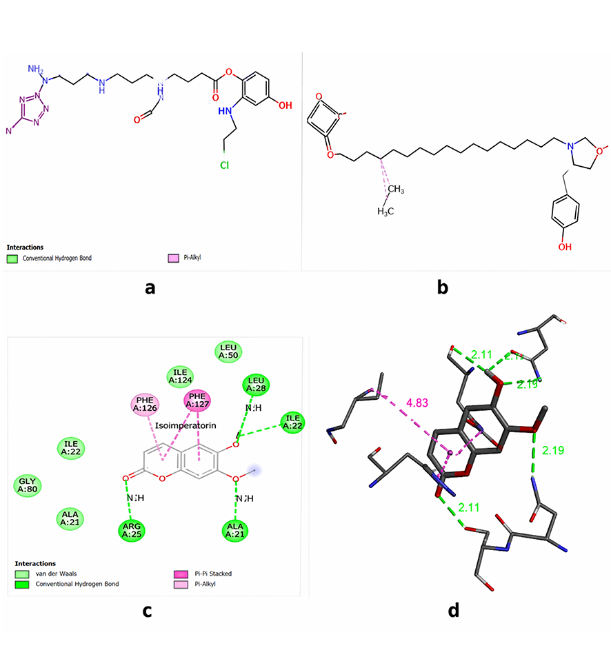
Figure 1. Comparison of isoscopoletin and 2-(4-hydroxyphenyl) ethyl lignocerate miglitol (standard inhibitor) in the active site of α-glucosidase enzyme
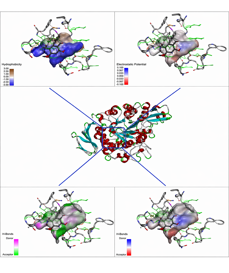
Figure 2. Two-dimensional (2D) and three-dimensional (3D) plots of α-glucosidase interactions with selected potential inhibitors
a) 2D representation of the molecular interactions between enzyme 2-(4-hydroxyphenyl) ethyl lignocerate b) 3D representation of molecular interactions between enzyme and 2-(4-hydroxyphenyl) ethyl lignocerate. c) 2D representation of molecular interactions between enzyme and isoscopoletin. d) 3D representation of molecular interactions between enzyme and isoscopoletin.
Figure 3. Three-dimensional representation of the receptor and isoscopoletin ligand surface interactions in the active site of α-glucosidase regarding polarity, hydrogen bonding, hydrophobic and ionized surface of the exchanges
Tables
Table 1. Evaluation of toxicity and toxicity class of the selected Ferula latisecta compounds.
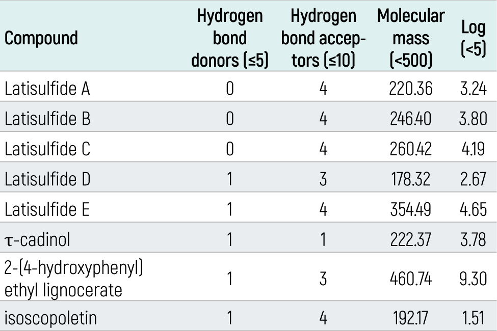
H: hepatotoxicity, NC: no carcinogenicity, NM: no mutagen, I: immunotoxicity
Table 2. Investigation of pharmacologic parameters (Drug-like) for sulfur compounds in F. latisecta according to Lipinski rule.
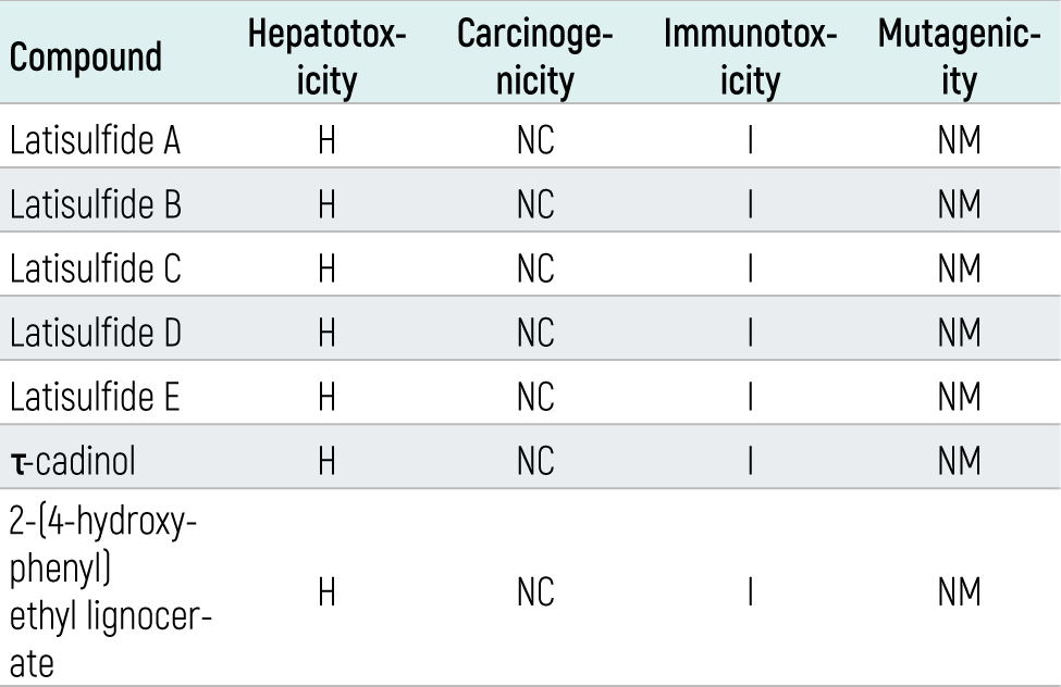
Table 3. Interaction and binding energy of sulfur compounds with amino acids of α-glucosidase.
Additional Information
Publisher’s Note
Bayrakol MP remains neutral with regard to jurisdictional and institutional claims.
Rights and Permissions

This work is licensed under a Creative Commons Attribution-NonCommercial 4.0 International License (CC BY-NC 4.0). To view a copy of the license, visit https://creativecommons.org/licenses/by-nc/4.0/
About This Article
How to Cite This Article
Sultan Mehtap Büyüker. In silico study of Ferula latisecta-derived compounds molecular interactions with α-glucosidase. Eu Clin Anal Med 2023;11(Suppl 1):S17-21. doi:10.4328/ECAM.10065
- Received:
- September 5, 2023
- Accepted:
- October 12, 2023
- Published Online:
- October 14, 2023
- Printed:
- October 15, 2023