Cardiac injury due to an utility knife hanging on the chest wall
Cardiac injury due to an utility knife
Authors
Penetrating thoracic traumas concerning heart or major vascular injuries can be life-threatening. In our case, we aimed to present a patient with cardiac injury, who has remarkable images.
Twenty-one year old male patient who is taking antipsychotic treatment had wounded himself in the anterior chest wall with an utility knife as attempting a suicide. Evaluation of the patient revealed a foreign body in the left hemithorax, left pleural effusion and pericardial effusion (Figure 1a).
Left thoracotomy was performed because of the sharp-edged foreign body which is greater than 1.5 cm in the neighborhood of the heart. A sharp foreign body protruding from the anterior chest wall (Figure 1b, 1c), hematoma in pericardial fatty tissue (Figure 1b), pericardial effusion and haemothorax was detected on the exploration of the thoracic cavity.
Following the removal of the foreign body, pericardial hematoma was evacuated by opening the pericardium. In the evaluation of myocardium, an injury approximately 1.5 cm in depth was detected millimeters away from the left anterior descending vein (Figure 1d). Damage was repaired with 6/0 pledged sutures (Figure 2). No complications had occurred in his follow-up and the patient was discharged afterwards.
Declarations
Animal and Human Rights Statement
All procedures performed in this study were in accordance with the ethical standards of the institutional and/or national research committee and with the 1964 Helsinki Declaration and its later amendments or comparable ethical standards.
Data Availability
The datasets used and/or analyzed during the current study are not publicly available due to patient privacy reasons but are available from the corresponding author on reasonable request.
Conflict of Interest
The authors declare that there is no conflict of interest.
Funding
None.
Scientific Responsibility Statement
The authors declare that they are responsible for the article’s scientific content, including study design, data collection, analysis and interpretation, writing, and some of the main line, or all of the preparation and scientific review of the contents, and approval of the final version of the article.
Figures
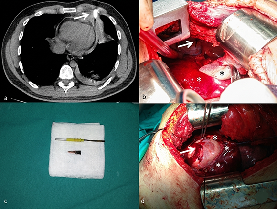
Figure 1. Thorax tomography of the patient showing the foreign body (a). Foreign body in the thoracic wall (arrow) and hematoma in the pericardial fatty pad (asteriks) (b). Part of the utility knife removed from the thoracic wall (c). Injury of the myocardium (arrow) which is seen after pericardiotomy (asterikses)(d).
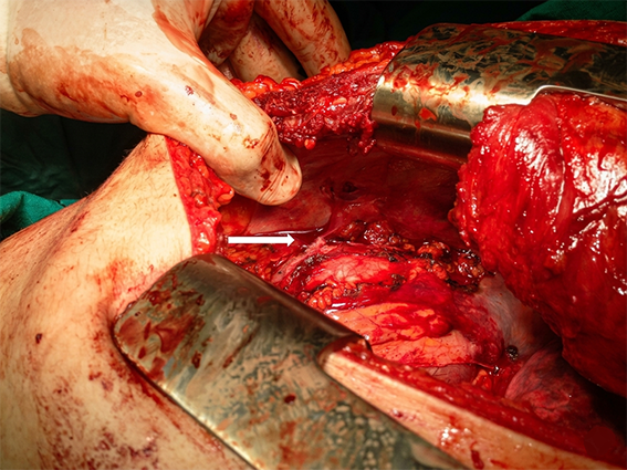
Figure 2. Repairment of the myocardium with pledget sutures (arrow).
Additional Information
Publisher’s Note
Bayrakol MP remains neutral with regard to jurisdictional and institutional claims.
Rights and Permissions

This work is licensed under a Creative Commons Attribution-NonCommercial 4.0 International License (CC BY-NC 4.0). To view a copy of the license, visit https://creativecommons.org/licenses/by-nc/4.0/
About This Article
How to Cite This Article
Sezai Çubuk, Okan Karataş, Cardiac Injury Due to an Utility Knife Hanging on the Chest Wall. Orhan Yücel. Eu Clin Anal Med 2013;1(2): doi:10.4328/ECAM.13
- Received:
- May 9, 2013
- Accepted:
- May 28, 2013
- Published Online:
- May 28, 2013
- Printed:
- May 28, 2013