An epidermal cyst mimicking a pilonidal cyst: A Case Report
An epidermal cyst mimicking a pilonidal cyst
Authors
Abstract
IntroductionEpidermal cysts consist of connective tissues including skin appendages coated by epithelia and the wall which may contain keratin, sebum, or follicle. Intergluteal sulcus is the specific region of localization for pilonidal cysts. Development of epidermal cysts in this region is extremely rare.Case PresentationWe present a 17-year-old woman with an epidermal cyst in the intergluteal region that mimicked a pilonidal cyst abscess. A 17-year-old woman presented to the emergency service with complaints of pain and swelling in the intergluteal region. A mass excision was performed. In the histopathological examination, the mass was reported an epidermal cyst.ConclusionThe possibility of an epidermal cyst should be kept in mind in the differential diagnosis of benign and malignant diseases of the perianal region.
Keywords
Introduction
Cystic masses can be located in any part of the body. These masses include epidermal cysts, which are also known as epidermoid, sebaceous, epithelial, and dermoid cysts. Epidermal cysts are mostly localized in the dermis that leads to increased epidermal thickness, resulting in the formation of stiff, elastic, and mobile masses. The cyst wall consists of connective tissues including skin appendages coated by epithelia and the wall which may contain keratin, sebum, or follicle.1 The central part of the cysts includes keratin-filled punctuation with a size of 5-50 mm. The cysts may grow with time and are rarely inflamed and painful, so the cyst may be confused with an abscess.In this report, we present a 17-year-old woman with an epidermal cyst in the intergluteal region that mimicked a pilonidal cyst abscess.
Case Presentation
A 17-year-old woman presented to the emergency service with complaints of pain and swelling in the intergluteal region. Patient history revealed that the patient had noticed the presence of stiffness in a small diameter of this region nine months previously, but had ignored the stiffness since there was no pain. Physical examination revealed a red subcutaneous mass in the intergluteal region with increased tenderness, which had a diameter of 3 cm (Figure 1). It was also revealed that the mass had been pre-diagnosed as perianal abscess and fistula by the pediatrics clinic and thus the association of the mass with the colon and rectum had been investigated via colonoscopy. No association had been found. Other system examinations were uneventful and the whole blood count and biochemical parameters were normal.Surface ultrasonography (USG) detected a 30x30 mm subcutaneous high-density hypoechoic mass. A magnetic resonance imaging (MRI) scan was performed to assess the proximity of the mass to other structures and MRI demonstrated a 34.7 x 30.1 mm hypodense mass (Figure 2).Mass excision was performed under spinal anesthesia. A Hemovac drain was inserted into the excision site. The excision site was closed using primary sutures. For prophylaxis, the patient was given 1 gr cefazolin sodium pre- and post-operatively. A postoperative analgesic, three doses of paracetamol, was provided subcutaneously. On postoperative day 2, the drain was removed and the patient was uneventfully discharged. On postoperative day 12, the patient was examined and the sutures were removed. In the histopathological examination, the mass was reported an epidermal cyst (Figure 3).
Discussion
Benign perianal masses are rare entities and are less common in men than in women. These masses are likely to grow and may rarely become infected and inflamed.2 The epidermal cysts in the perianal region tend to appear on the surface of the skin and tend to be yellowish in color. The differential diagnosis of perianal cysts includes hemorrhoids, fistulas, abscesses, pilonidal sinus/cysts, perianal dermatosis, anal duct cysts, benign teratomas, epidermal and dermoid cysts, perianal skin cancers, and malign teratomas.1 The abscess formations in the intergluteal region usually result from infection in pilonidal sinus. In this region, perianal fistulas and abscesses may also be seen, though rarely. To date, the literature has not reported an epidermal cyst localized in this region. The cases reported in the literature are mainly localized in the scrotum, perineum, labium, and gluteus.3,4 Epidermal cysts are commonly treated by total excision, whereas pilonidal abscesses are treated by drainage. In addition, although antibiotic therapy is required following the drainage of abscesses, no antibiotic therapy is needed following the excision of epidermal cysts.Laboratory tests have no diagnostic value in the diagnosis of cysts. In our case, the laboratory parameters were within normal range. USG, MRI, and computed tomography (CT) are useful in the clinical diagnosis of cystic masses. In our case, USG was performed due to the pre-diagnosis of pilonidal sinus abscess and pelvic MRI was performed because of the diagnosis of a high-density mass. USG provides useful outcomes in the differentiation of cysts from solid tumors, whereas MRI is a superior tool in assessing the proximity of masses to other structures. On the other hand, colonoscopy may be useful for avoiding missing other accompanying lesions. In our case, the diagnosis of perianal fistula was ruled out by colonoscopy. Nevertheless, despite all of these available tests, the pathologies localized in this region can only be definitively diagnosed by a histopathologic analysis.Keeping the possibility of epidermoid cysts in mind in the differential diagnosis of intergluteal masses and abscesses is of prime importance for the administration of a suitable antibiotic therapy and proper surgical treatment.
Declarations
Animal and Human Rights Statement
All procedures performed in this study were in accordance with the ethical standards of the institutional and/or national research committee and with the 1964 Helsinki Declaration and its later amendments.
Informed Consent
Informed consent was obtained from all participants.
Conflict of Interest
The authors declare no conflicts of interest.
Funding
None.
Scientific Responsibility Statement
The authors declare that they are responsible for the scientific content of the article, including the study design, data collection, analysis and interpretation, manuscript preparation, and approval of the final version of the manuscript.
References
-
Ramaswamy AS, Manjunatha HK, Sunilkumar B, Arunkumar SP. Morphological spectrum of pilar cysts. N Am J Med Sci. 2013;5(2):124-128. doi:10.4103/1947-2714.107532
-
Abreu Velez AM, Brown VM, Howard MS. An inflamed trichilemmal (pilar) cyst: not so simple? N Am J Med Sci. 2011;3:431-434. doi:10.4297/najms.2011.3431
-
Karaca S, Kulaç M, Dilek FH, Polat C, et al. Giant proliferating trichilemmal tumor of the gluteal region. Dermatol Surg. 2005;31(12):1734-1736. doi:10.2310/6350.2005.31323
-
Machida T, Matsuoka Y, Kobayashi S, Ozeki Z, Ishizaka K, Oka T. Case of giant perineal epidermal cyst: a case report. Hinyokika Kiyo. 2003;49:257-259.
Figures
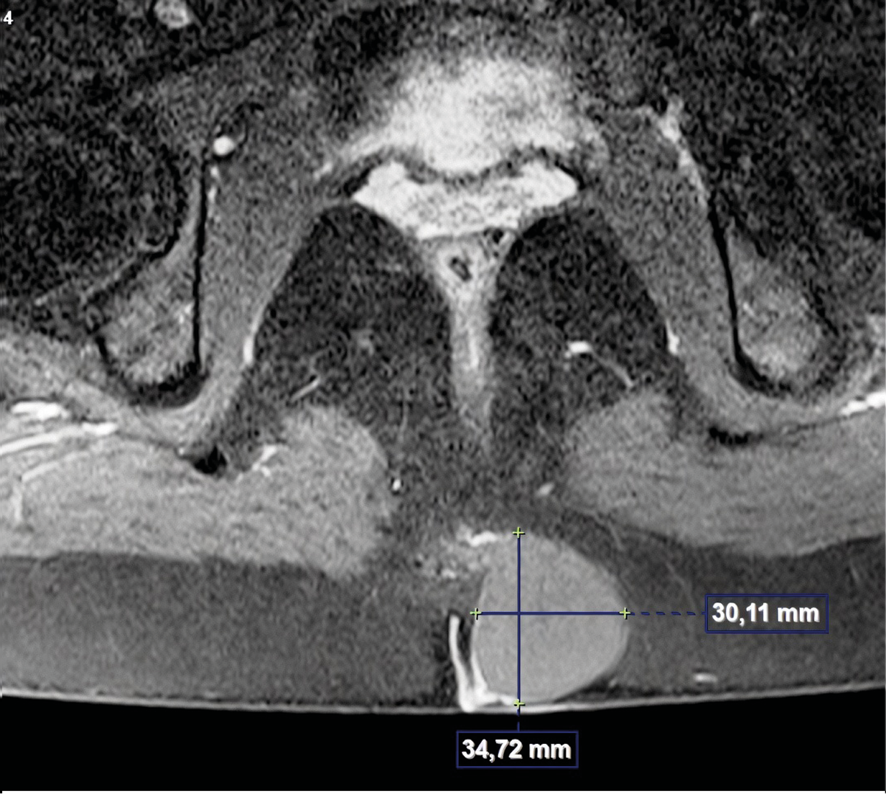
Figure 1. Computed tomography image of epidermal cyst
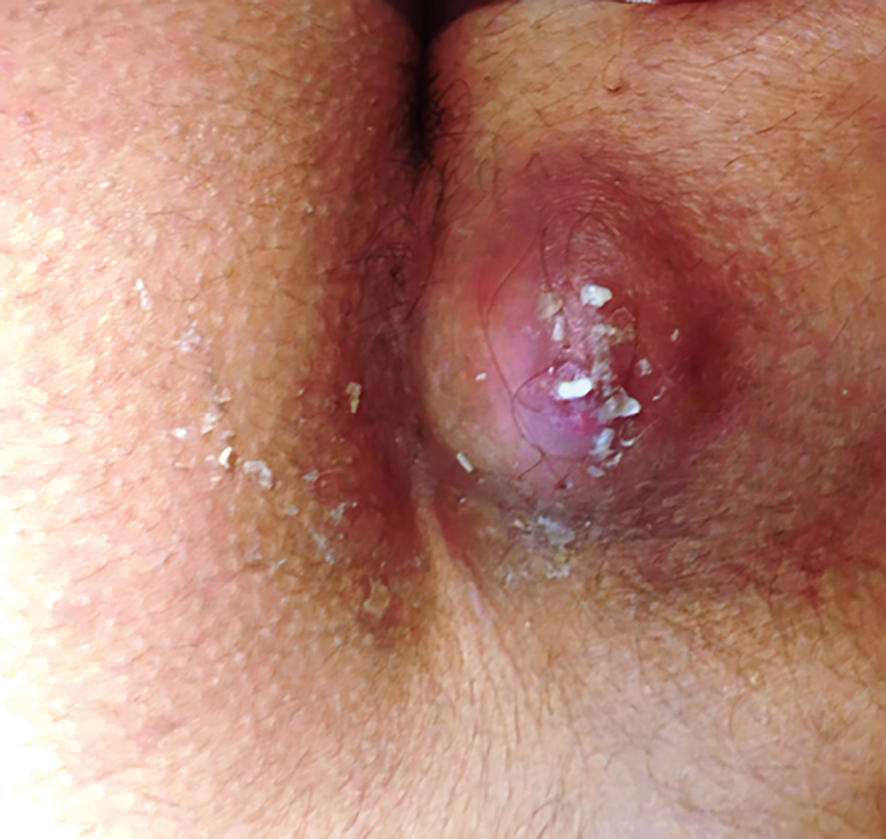
Figure 2. Epidermal cyst

Figure 3. Histopathology of epidermal cyst
Additional Information
Publisher’s Note
Bayrakol MP remains neutral with regard to jurisdictional and institutional claims.
This study was accepted as a poster presentation in the 10th Congress of Turkish Traumatology and Emergeny, 2015 in Antalya, Turkey.
Rights and Permissions

This work is licensed under a Creative Commons Attribution-NonCommercial 4.0 International License (CC BY-NC 4.0). To view a copy of the license, visit https://creativecommons.org/licenses/by-nc/4.0/
About This Article
How to Cite This Article
Yavuz Savas Koca, İhsan Yıldız, Mustafa Ugur, Tugba Gürsoy Koca. An epidermal cyst mimicking a pilonidal cyst: A Case Report. Eu Clin Anal Med 2017;5(1):14-16. doi:10.4328/ECAM.103
- Received:
- November 14, 2016
- Accepted:
- December 2, 2016
- Published Online:
- January 1, 2017
- Printed:
- January 1, 2017